Zurück

Testen Sie Ihre IntelliVence (Band 15, Ausgabe 4)

Artikel

Autor: Clinical Experts Group, Hamilton Medical Group

Datum: 08.08.2018

Warum unterscheidet sich der aktuelle Loop für das volumetrische Kapnogramm vom Referenz‑Loop?

Testen Sie Ihre IntelliVence (Band 15, Ausgabe 4)

Antwort

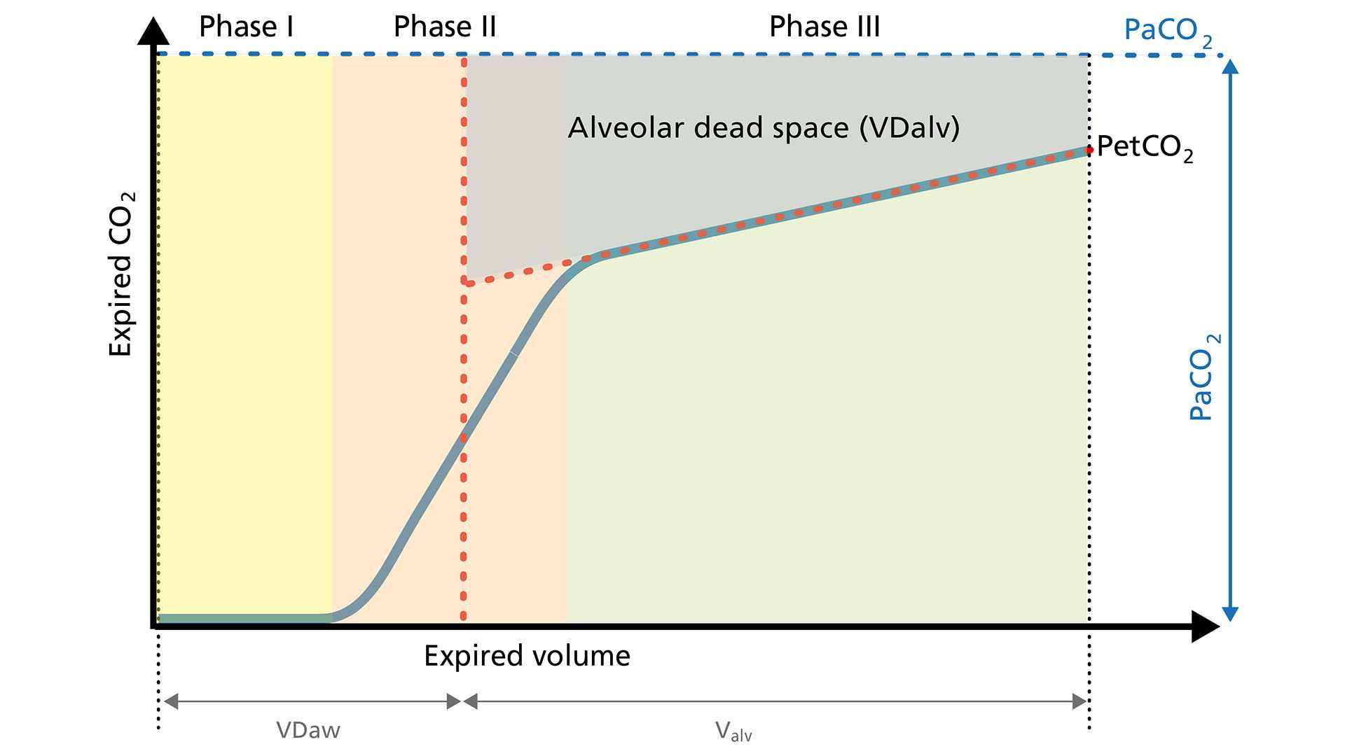
Lungenembolie (LE). Bei einer Lungenembolie ist der alveoläre Totraum (VDalv) durch den verringerten Blutfluss in die gut belüfteten Alveolen erhöht. Dieses Gas mit niedrigem CO2‑Gehalt wird synchron mit dem Gas aus normal perfundierten Alveolen ausgeatmet. Darin unterscheidet sich die LE von Lungenerkrankungen, die den Atemweg betreffen und die durch eine asynchrone Entleerung der Lungenkompartimente mit einem ungleichmäßigen Ventilations‑Perfusions‑Verhältnis gekennzeichnet sind (Eriksson L, Wollmer P, Olsson CG, et al. Diagnosis of pulmonary embolism based upon alveolar dead space analysis. Chest. 1989;96(2):357-362. doi:10.1378/chest.96.2.3571​). 
 

Repräsentative Darstellung eines Kapnogramms mit drei Phasen und einem flacheren Plateau in Phase III
Normales volumetrisches Kapnogramm mit Phase I, II und III sowie VDalv

Mit dem Anstieg von VDalv nimmt der CO2‑Gehalt im ausgeatmeten Gas ab, was in weiterer Folge zu einem Abfallen des VCO2‑Wertes führt. Der Loop für das volumetrische Kapnogramm ist durch einen deutlich niedrigeren PetCO2‑Wert und ein flacheres Plateau in Phase III gekennzeichnet. Außerdem ist der Anstieg in Phase II weniger steil.

Repräsentative Darstellung eines Kapnogramms mit drei Phasen und einem flacheren Plateau in Phase III
Normales volumetrisches Kapnogramm mit Phase I, II und III sowie VDalv

Diagnosis of pulmonary embolism based upon alveolar dead space analysis.

Eriksson L, Wollmer P, Olsson CG, et al. Diagnosis of pulmonary embolism based upon alveolar dead space analysis. Chest. 1989;96(2):357‑362. doi:10.1378/chest.96.2.357

Pulmonary embolism (PE) leads to an abnormal alveolar deadspace that is expired in synchrony with gas from normally perfused alveoli. This feature of PE separates it from pulmonary diseases affecting the airways, which are characterized by nonsynchronous emptying of compartments with an uneven ventilation/perfusion relationship. An analysis of the single breath test (SBT) for CO2, SBT‑CO2, focusing on the late tidal expirate, was made in order to evaluate the feasibility to use the SBT‑CO2 for the diagnosis of PE. The test was evaluated in 38 patients with suspected PE where pulmonary angiography showed that nine had PE and 29 did not. It was also tested in a reference population consisting of patients with normal lung function, obstructive lung disease and interstitial lung disease. Previously suggested gas exchange measurements for the diagnosis of PE, ie, the physiologic deadspace fraction, VDphys/VT, and the arterial‑to‑end‑tidal CO2 gradient, P(a‑E')CO2, were also evaluated in the groups. SBT‑CO2 achieved a nearly complete separation between the patients with PE and those without. The other measurements, however, showed a substantial overlap between patients with PE and those with obstructive or interstitial lung disease. The SBT‑CO2 is simple and potentially widely available and warrants further study as a routine technique for the diagnosis of PE.