Zurück

Messung von AutoPEEP und gesamtem PEEP

Artikel

Autor: Clinical Experts Group, Hamilton Medical

Datum: 14.07.2017

Last change: 30.09.2020

(Originally published 14.07.2017) Previously: select Exp hold, when flow=0 select Exp hold again to deactivate hold maneuver. SW versions updated.

Im Falle einer dynamischen Hyperinflation der Lunge übersteigt der durchschnittliche endexspiratorische Druck in den Alveolen (also der tatsächliche gesamte PEEP (PEEPtot)) den durch das Beatmungsgerät angewendeten PEEP-Wert (PEEPe). Die Differenz zwischen PEEPtot und PEEPe entspricht dem intrinsischen PEEP (PEEPi); sie wird auch AutoPEEP genannt (1).

Messung von AutoPEEP und gesamtem PEEP

AutoPEEP und RCexsp

AutoPEEP wird auch als Airtrapping, Breath Stacking, dynamische Hyperinflation, unbeabsichtigter PEEP oder versteckter PEEP bezeichnet.

AutoPEEP ist ein bekanntes Phänomen bei maschinell beatmeten Patienten mit langen exspiratorischen Zeitkonstanten (RCexsp), z. B. bei Patienten mit chronischer obstruktiver Lungenerkrankung (COPD) oder akutem starkem Asthma.

WICHTIG: Der resultierende AutoPEEP-Wert ist nicht auf der Kurve für den Atemwegsdruck sichtbar, die während der normalen Verabreichung des Atemhubs auf dem Bildschirm des Beatmungsgerätes angezeigt wird.

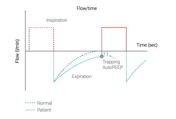
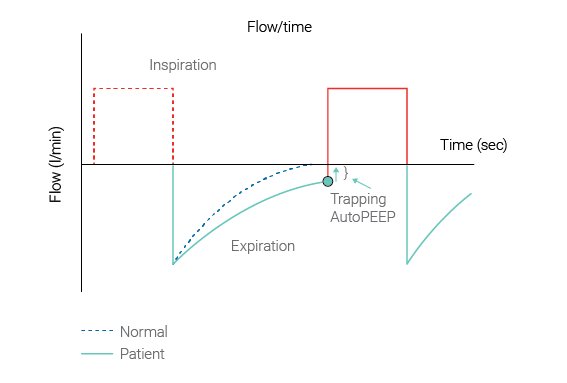
(Abbildung 1 unten: Quelle Garcia Vicente et al. (García Vicente E, Sandoval Almengor JC, Díaz Caballero LA, Salgado Campo JC. Ventilación mecánica invasiva en EPOC y asma [Invasive mechanical ventilation in COPD and asthma]. Med Intensiva. 2011;35(5):288-298. doi:10.1016/j.medin.2010.11.0042​))

Flow-/Zeit-Grafik mit AutoPEEP und Airtrapping
Abbildung 1: AutoPEEP und Airtrapping
Flow-/Zeit-Grafik mit AutoPEEP und Airtrapping
Abbildung 1: AutoPEEP und Airtrapping

Auswirkungen von AutoPEEP

AutoPEEP ist der Grund für die Veranlagung eines Patienten zu erhöhter Atemarbeit, Barotrauma, hämodynamischer Instabilität und Schwierigkeiten bei der Triggerung des Beatmungsgerätes. Wenn die hämodynamischen Auswirkungen von AutoPEEP nicht erkannt werden, kann dies zu einer unangemessenen Beschränkung der Flüssigkeiten oder einer nicht notwendigen Therapie mit Vasopressoren führen. AutoPEEP kann die Entwöhnung von der maschinellen Beatmung behindern.

Pflegekräfte sollten auf das Auftreten von AutoPEEP während der Beatmung achten und die Parameter zur Beatmungskontrolle entsprechend einstellen, um die negativen Auswirkungen von PEEP zu vermeiden.

AutoPEEP berechnen

Alle Beatmungsgeräte von Hamilton Medical sind mit der einzigartigen Fähigkeit ausgestattet, AutoPEEP als Monitoring-Parameter Atemhub für Atemhub anzuzeigen. Er wird anhand der Methode der kleinsten Quadrate (LSF) über den gesamten Atemhub berechnet (Iotti GA, Braschi A, Brunner JX, et al. Respiratory mechanics by least squares fitting in mechanically ventilated patients: applications during paralysis and during pressure support ventilation. Intensive Care Med. 1995;21(5):406-413. doi:10.1007/BF017074093​). Unter bestimmten Umständen, z. B. im Falle einer starken dynamischen Hyperinflation, kann jedoch der mit der LSF-Methode berechnete AutoPEEP unter dem tatsächlichen AutoPEEP liegen. In solchen Fällen kann der genaue Wert mithilfe eines exspiratorischen Hold-Manövers ermittelt werden.

So messen Sie den gesamten PEEP mithilfe eines exspiratorischen Hold-Manövers (siehe Abbildung 2 unten):

Stellen Sie sicher, dass die Paw-Kurve angezeigt wird.

  1. Öffnen Sie das Fenster Hold.
  2. Warten Sie, bis die Anzeige der Paw-Kurve an der linken Seite wieder gestartet wird.
  3. Warten Sie auf die nächste Inspiration.
  4. Wählen Sie dann Hold Exsp. Warten Sie 3 bis 5 Sekunden, wählen Sie dann Hold Exsp oder drücken Sie den Einstellknopf erneut, um das Hold-Manöver zu deaktivieren, und schliessen Sie das Fenster.
  5. Nach dem Manöver wird das Fenster „Hold“ geschlossen und die Funktion „Einfrieren“ wird automatisch aktiviert.
  6. Ermitteln Sie den gesamten PEEP, indem Sie mit dem Cursor auf der Druckkurve die Punkte hinter der Stelle anzeigen, wo der Flow null erreicht hat.
  7. Berechnen Sie AutoPEEP, indem Sie den extrinsischen PEEP vom gesamten PEEP abziehen.

Berechnungen

AutoPEEP '= gesamter PEEP - extrinsischer PEEP = intrinsischer PEEP
PEEP '= extrinsischer PEEP und ist vorausgewählt
Gesamter PEEP '= intrinsischer PEEP + extrinsischer PEEP
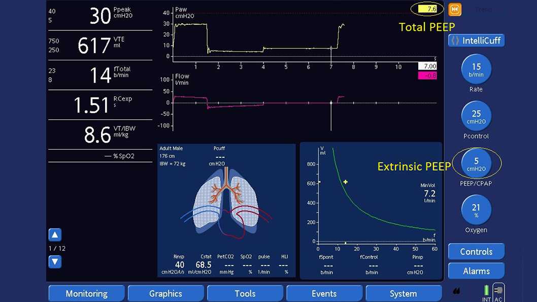
Screenshot des Displays des Beatmungsgerätes mit Anzeige eines exspiratorischen Hold-Manövers
Abbildung 2: Gesamter PEEP wird mithilfe eines exspiratorischen Hold-Manövers gemessen. Gesamter PEEP von 7,6 cmH2O - extrinsischer PEEP von 5 cmH2O = AutoPEEP von 2,6 cmH2O
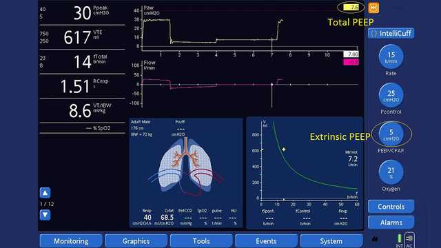
Screenshot des Displays des Beatmungsgerätes mit Anzeige eines exspiratorischen Hold-Manövers
Abbildung 2: Gesamter PEEP wird mithilfe eines exspiratorischen Hold-Manövers gemessen. Gesamter PEEP von 7,6 cmH2O - extrinsischer PEEP von 5 cmH2O = AutoPEEP von 2,6 cmH2O

Airtrapping vermeiden

Wenn ein unbeabsichtigter AutoPEEP vorhanden ist, sollten Pflegekräfte eine Anpassung der Kontrollparameter erwägen, um durch eine Erhöhung der Exspirationszeit Airtrapping zu vermeiden. Die Verwendung von Endotrachealtuben mit grossem Durchmesser oder von Bronchodilatatoren, eine kurze Einstellung für die Inspirationszeit bzw. eine lange Einstellung für die Exspirationszeit, niedrigere Atemfrequenzen und der Einsatz von Beruhigungsmitteln können nötig sein, um eine dynamische Hyperinflation aufgrund von Airtrapping zu vermeiden.

Jedes Beatmungsgerät von Hamilton Medical verfügt über den intelligenten Beatmungsmodus Adaptive Support Ventilation (ASV®). ASV verwendet automatisch Lungenschutzstrategien, um Komplikationen wie AutoPEEP zu vermindern.

Betroffene Geräte: HAMILTON-G5/S1 (ab SW-Version 2.8x); HAMILTON-C3 (ab SW-Version 2.0.x), HAMILTON-C6 (ab SW-Version 1.1.x)

Den vollständigen Quellenverweis finden Sie unten (Iotti, G., & Braschi, A. (1999). Measurements of respiratory mechanics during mechanical ventilation. Rhäzüns, Switzerland: Hamilton Medical Scientific Library.1​).

Measurements of respiratory mechanics during mechanical ventilation

Iotti, G., & Braschi, A. (1999). Measurements of respiratory mechanics during mechanical ventilation. Rhäzüns, Switzerland: Hamilton Medical Scientific Library.

Invasive mechanical ventilation in COPD and asthma.

García Vicente E, Sandoval Almengor JC, Díaz Caballero LA, Salgado Campo JC. Ventilación mecánica invasiva en EPOC y asma [Invasive mechanical ventilation in COPD and asthma]. Med Intensiva. 2011;35(5):288-298. doi:10.1016/j.medin.2010.11.004

COPD and asthmatic patients use a substantial proportion of mechanical ventilation in the ICU, and their overall mortality with ventilatory support can be significant. From the pathophysiological standpoint, they have increased airway resistance, pulmonary hyperinflation, and high pulmonary dead space, leading to increased work of breathing. If ventilatory demand exceeds work output of the respiratory muscles, acute respiratory failure follows. The main goal of mechanical ventilation in this kind of patients is to improve pulmonary gas exchange and to allow for sufficient rest of compromised respiratory muscles to recover from the fatigued state. The current evidence supports the use of noninvasive positive-pressure ventilation for these patients (especially in COPD), but invasive ventilation also is required frequently in patients who have more severe disease. The physician must be cautious to avoid complications related to mechanical ventilation during ventilatory support. One major cause of the morbidity and mortality arising during mechanical ventilation in these patients is excessive dynamic pulmonary hyperinflation (DH) with intrinsic positive end-expiratory pressure (intrinsic PEEP or auto-PEEP). The purpose of this article is to provide a concise update of the most relevant aspects for the optimal ventilatory management in these patients.

Respiratory mechanics by least squares fitting in mechanically ventilated patients: applications during paralysis and during pressure support ventilation.

Iotti GA, Braschi A, Brunner JX, et al. Respiratory mechanics by least squares fitting in mechanically ventilated patients: applications during paralysis and during pressure support ventilation. Intensive Care Med. 1995;21(5):406-413. doi:10.1007/BF01707409


OBJECTIVE

To evaluate a least squares fitting technique for the purpose of measuring total respiratory compliance (Crs) and resistance (Rrs) in patients submitted to partial ventilatory support, without the need for esophageal pressure measurement.

DESIGN

Prospective, randomized study.

SETTING

A general ICU of a University Hospital.

PATIENTS

11 patients in acute respiratory failure, intubated and assisted by pressure support ventilation (PSV).

INTERVENTIONS

Patients were ventilated at 4 different levels of pressure support. At the end of the study, they were paralyzed for diagnostic reasons and submitted to volume controlled ventilation (CMV).

MEASUREMENTS AND RESULTS

A least squares fitting (LSF) method was applied to measure Crs and Rrs at different levels of pressure support as well as in CMV. Crs and Rrs calculated by the LSF method were compared to reference values which were obtained in PSV by measurement of esophageal pressure, and in CMV by the application of the constant flow, end-inspiratory occlusion method. Inspiratory activity was measured by P0.1. In CMV, Crs and Rrs measured by the LSF method are close to quasistatic compliance (-1.5 +/- 1.5 ml/cmH2O) and to the mean value of minimum and maximum end-inspiratory resistance (+0.9 +/- 2.5 cmH2O/(l/s)). Applied during PSV, the LSF method leads to gross underestimation of Rrs (-10.4 +/- 2.3 cmH2O/(l/s)) and overestimation of Crs (+35.2 +/- 33 ml/cmH2O) whenever the set pressure support level is low and the activity of the respiratory muscles is high (P0.1 was 4.6 +/- 3.1 cmH2O). However, satisfactory estimations of Crs and Rrs by the LSF method were obtained at increased pressure support levels, resulting in a mean error of -0.4 +/- 6 ml/cmH2O and -2.8 +/- 1.5 cmH2O/(l/s), respectively. This condition was coincident with a P0.1 of 1.6 +/- 0.7 cmH2O.

CONCLUSION

The LSF method allows non-invasive evaluation of respiratory mechanics during PSV, provided that a near-relaxation condition is obtained by means of an adequately increased pressure support level. The measurement of P0.1 may be helpful for titrating the pressure support in order to obtain the condition of near-relaxation.