Back

Using volumetric capnography to set PEEP

Article

Author: Jean-Michel Arnal, Senior Intensivist, Ste Musse Hospital, Toulon, France

Date of first publication: 23.04.2019

PEEP is used to keep the lung aerated and prevent lung collapse at the end of expiration. However, PEEP may over-distend the normally aerated lung and impair lung perfusion. Therefore, any change in PEEP may affect the overall ventilation/perfusion ratio in an unpredictable way.

Using volumetric capnography to set PEEP

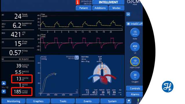
Volumetric capnography measures the volume of CO2 exhaled breath-by-breath (VeCO2). After a change in PEEP, assuming that the cardiovascular function and tidal volume are stable, an increase in VeCO2 means that the overall ventilation/perfusion ratio has improved. Conversely, a decrease in VeCO2 means that the ventilation/perfusion ratio is worsening. VeCO2 changes rapidly and returns to the baseline after a few minutes.

The limitation to this method is that the clinician monitors rapid changes to the ventilation/perfusion ratio, such as those due to lung over-distension and lung-perfusion impairment or improvement.

After a change in PEEP, recruitment or derecruitment may take a longer time to occur and cannot be assessed by this method.

Watch the video below to see a demonstration during ventilation with a Hamilton Medical ventilator. 


Full citations below: (Blankman P, Shono A, Hermans BJ, Wesselius T, Hasan D, Gommers D. Detection of optimal PEEP for equal distribution of tidal volume by volumetric capnography and electrical impedance tomography during decreasing levels of PEEP in post cardiac-surgery patients. Br J Anaesth. 2016;116(6):862-869. doi:10.1093/bja/aew1161​)

Volumetric Capnography: How to set PEEP according to VCO2

Senior intensivist Dr. Jean-Michel Arnal demonstrates how volumetric capnography can help to find the correct PEEP setting.
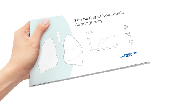
Volumentric Capnography e-book

Free e-book

Good to know! All about volumetric capnography

Learn how to interpret a volumetric capnogram and get an overview of the benefits and clinical applications of volumetric capnography. Includes a self-test!

Detection of optimal PEEP for equal distribution of tidal volume by volumetric capnography and electrical impedance tomography during decreasing levels of PEEP in post cardiac-surgery patients.

Blankman P, Shono A, Hermans BJ, Wesselius T, Hasan D, Gommers D. Detection of optimal PEEP for equal distribution of tidal volume by volumetric capnography and electrical impedance tomography during decreasing levels of PEEP in post cardiac-surgery patients. Br J Anaesth. 2016;116(6):862-869. doi:10.1093/bja/aew116


BACKGROUND

Homogeneous ventilation is important for prevention of ventilator-induced lung injury. Electrical impedance tomography (EIT) has been used to identify optimal PEEP by detection of homogenous ventilation in non-dependent and dependent lung regions. We aimed to compare the ability of volumetric capnography and EIT in detecting homogenous ventilation between these lung regions.

METHODS

Fifteen mechanically-ventilated patients after cardiac surgery were studied. Ventilator settings were adjusted to volume-controlled mode with a fixed tidal volume (Vt) of 6-8 ml kg(-1) predicted body weight. Different PEEP levels were applied (14 to 0 cm H2O, in steps of 2 cm H2O) and blood gases, Vcap and EIT were measured.

RESULTS

Tidal impedance variation of the non-dependent region was highest at 6 cm H2O PEEP, and decreased significantly at 14 cm H2O PEEP indicating decrease in the fraction of Vt in this region. At 12 cm H2O PEEP, homogenous ventilation was seen between both lung regions. Bohr and Enghoff dead space calculations decreased from a PEEP of 10 cm H2O. Alveolar dead space divided by alveolar Vt decreased at PEEP levels ≤6 cm H2O. The normalized slope of phase III significantly changed at PEEP levels ≤4 cm H2O. Airway dead space was higher at higher PEEP levels and decreased at the lower PEEP levels.

CONCLUSIONS

In postoperative cardiac patients, calculated dead space agreed well with EIT to detect the optimal PEEP for an equal distribution of inspired volume, amongst non-dependent and dependent lung regions. Airway dead space reduces at decreasing PEEP levels.