We have detected that you are visiting our website from United States.
We offer a separate version of the website for your country (United States).

Switch to United States
 Products

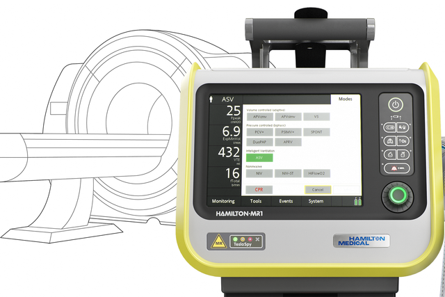
HAMILTON-MR1. Intelligent Ventilation from the ICU to MRI

HAMILTON-MR1_MRI_desktop_01

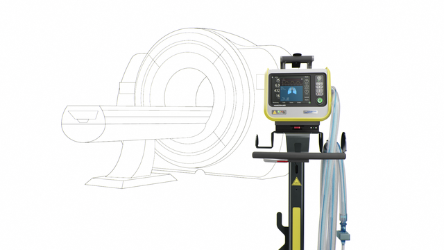
Our MRI specialist. For high-end ventilation during MRI procedures

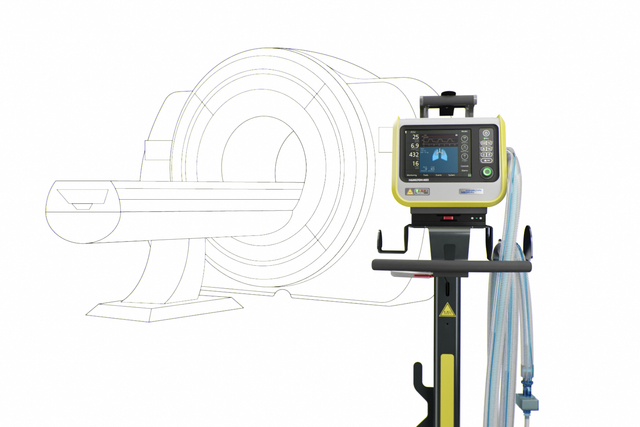
  • Comprehensive ICU ventilator for use in the MRI department
  • MR-Conditional with the use of 1.5 Tesla and 3.0 Tesla static magnetic field scanners
  • The ventilator can remain close to the patient even during MRI scans
  • For all patient populations
HAMILTON-MR1_MRI_desktop_01
HAMILTON-MR1

Our MRI specialist. For high-end ventilation during MRI procedures
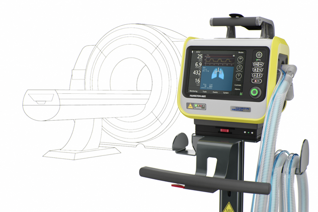
  • Comprehensive ICU ventilator for use in the MRI department
  • MR-Conditional with the use of 1.5 Tesla and 3.0 Tesla static magnetic field scanners
  • The ventilator can remain close to the patient even during MRI scans
  • For all patient populations
HAMILTON-MR1

Walk with me! From the patient room to the MRI suite

  • Up to 540 minutes of battery time
  • High-performance turbine
  • Compact and portable
HAMILTON-MR1

Continuous ventilation therapy. Same mode and same settings as at the bedside

  • Volume‑controlled and pressure‑controlled ventilation modes
  • Adaptive ventilation with ASV®
  • Noninvasive ventilation
  • High flow nasal cannula therapy
HAMILTON-MR1

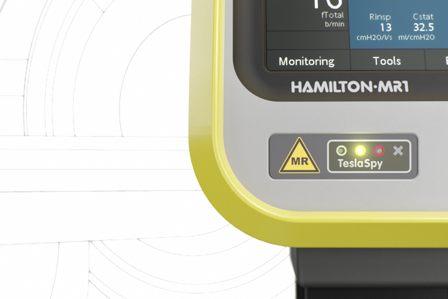
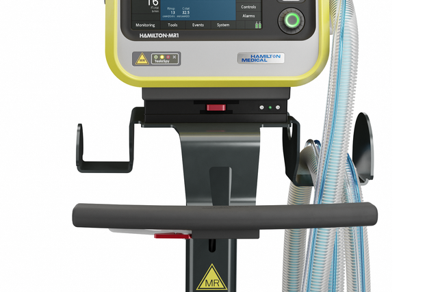
Always on the lookout. Your magnetic field navigator

The TeslaSpy helps you to keep the ventilator at a safe distance from the MRI scanner.
HAMILTON-MR1

Two strong arms. For the extra-long breathing circuits

Choose between a range of breathing circuits in different lengths suitable for the MRI suite.
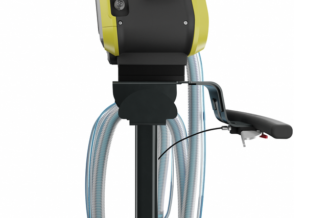
HAMILTON-MR1

Stay! Locked in place automatically

The auto-lock brakes on the trolley automatically lock in place, preventing the ventilator from accidentally rolling toward the MRI scanner.
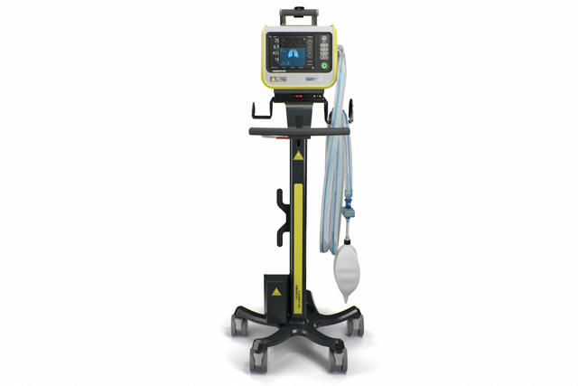
HAMILTON-MR1
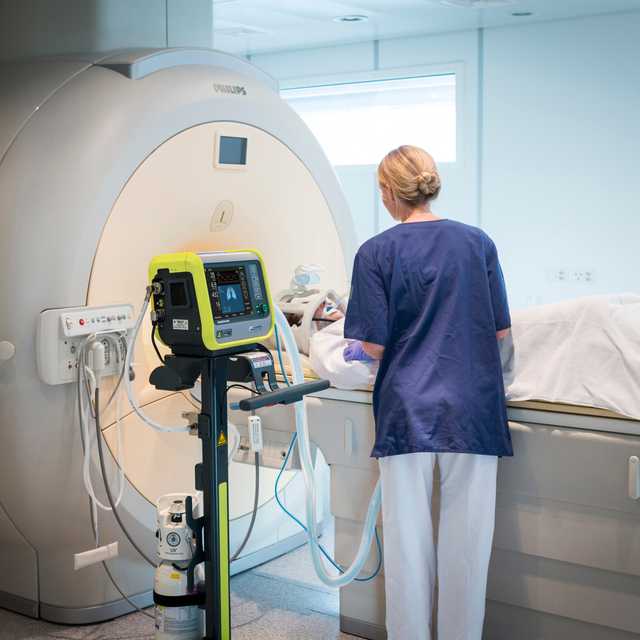
HAMILTON-MR1; use during MRI scan

Come closer! MR-Conditional up to 50 mT

The HAMILTON-MR1 allows you to get close to the MRI scanner, because it is designed to withstand a static magnetic field of up to 50 mT. In comparison to ventilators with weaker shielding from the magnetic field, it gives you more freedom when positioning the device, a wider range of movement, and greater flexibility for your workflow and patient setup.

Closer proximity to the scanner also means you can use shorter breathing circuits. The advantage is their smaller compressible volume, which can contribute to better overall ventilation quality.
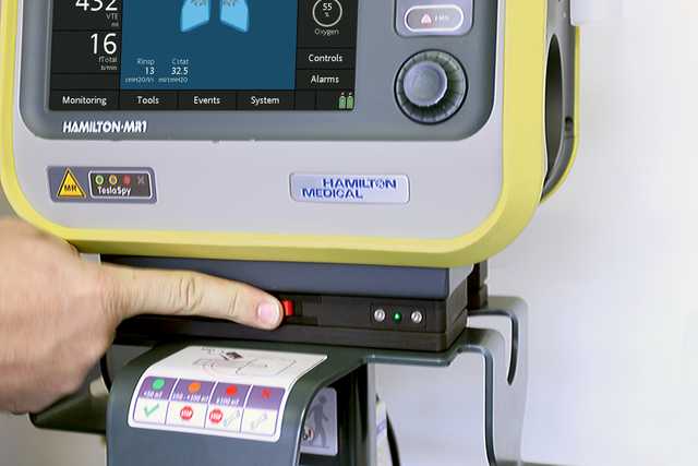
HAMILTON-MR1 pushing quick release button

Click on, click off. For an easy transition

The quick‑release button on the transport kit allows you to detach the device from the trolley with just the push of a button. And it reattaches just as easily.

With the new Software update 3.0 for the HAMILTON-MR1, the audiovisual TeslaSpy alarms on the are now mirrored on both the ventilator display and the alarm lamp for greater visibility. TeslaSpy is a magnetic field navigator which continuously measures the background magnetic levels, even when the ventilator is switched off. It lets you know when magnetic field levels are safe, and when they exceed the ventilator’s safety threshold. The TeslaSpy alarms are now also included in the Event log. The Software Update 3.0 brings a lot of new features to our compact devices. To learn more go to: www.hamilton-medical.com/compact-device-more-features The availability of SW v3.0.x, as well as individual features, depends on the specific device and market.

Your lookout is on board. To navigate the magnetic field

The onboard magnetic field navigator, TeslaSpy, continuously monitors the magnetic field and gives you an audible and visual signal if you are getting too close. Positioning a medical device too close to the MRI scanner can have fatal consequences.

The TeslaSpy alarms are mirrored on the device's GUI and on the alarm lamp. For maximum safety, TeslaSpy continues monitoring even when the ventilator is switched off.

Want to see more?
Explore the 3D model

Discover the HAMILTON-MR1 from every angle and click on the hotspots to learn more.

For quick details

  • Standard
  • Option
  • Not available
Patient groups Adult/Ped, Neonatal
Dimensions (W x D x H) 320 x 220 x 270 mm (ventilation unit)
630 x 630 x 1400 mm (incl. trolley)
Weight 6.8 kg (15 lb)
21 kg (46.2 lb) with trolley
Monitor size and resolution 8.4 in (214 mm) diagonal
640 x 480 pixels
Detachable monitor
Battery operating time 8 h with two batteries
Hot-swappable battery
Air supply Integrated turbine
O2 connector DISS (CGA 1240) or NIST
Connectivity USB port
Loudness 42 dB in normal operation
Volume controlled, flow controlled
Volume targeted, adaptive pressure controlled
Intelligent ventilation ASV®
Noninvasive ventilation
High flow
Visualization of lung mechanics (Dynamic Lung)
Visualization of the patient’s ventilator dependence
Esophageal pressure measurement
Capnography
SpO2 monitoring
Recruitability assessment and lung recruitment (P/V Tool Pro)
Patient-ventilator synchronization (IntelliSync+)
CPR ventilation
Hamilton Connect Module
Remote connection to HAMILTON-H900 humidifier
Integrated IntelliCuff cuff pressure controller
Integrated pneumatic nebulizer
Integrated Aerogen nebulizer
Compatibility with Sedaconda ACD-S anesthetic delivery system
Dr. Adrian Wäckerlin Thomas Berlin

Customer voices

Until now, we ventilated our ICU patients during MRI scanning with an anesthesia ventilator. Therefore, we had to consult an anesthetist every time to handle the equipment. With the HAMILTON-MR1, we are now completely independent.

Dr. Adrian Wäckerlin

Head of ICU
Grisons Cantonal Hospital, Chur, Switzerland

Customer voices

You can take the patient on the HAMILTON‑MR1 from the ICU down to the magnetic resonance imaging suite for an MR study and not have to change a thing about the mechanical ventilation. And that is a true advantage because you are not risking lung de‑recruitment and a patient setback, which would keep the patient in the hospital longer and make it more uncomfortable for them.

Thomas Berlin

Director of Respiratory Care
AdventHealth Orlando, Orlando (FL), USA

For your patients

Intelligent ventilation solutions at a glance

ASV® - Adaptive Support Ventilation®. For adaptation around the clock

The ventilation mode ASV continuously adjusts the respiratory rate, tidal volume, and inspiratory time breath by breath depending on the patient’s lung mechanics and effort - 24 hours a day, from intubation to extubation.

Integrated nebulizer. For additional treatments

The integrated pneumatic nebulizer is fully synchronized with the timing of inspiration and expiration.

An integrated, synchronized Aerogen nebulizer is available as an option (Not available in all marketsa​, Only available for HAMILTON-C6/G5/S1b​).

The delivery of a fine mist of drug aerosol particles helps you reverse bronchospasm, improve ventilation efficiency, and reduce hypercapnia (Dhand R. New frontiers in aerosol delivery during mechanical ventilation. Respir Care. 2004;49(6):666-677. 100​, Waldrep JC, Dhand R. Advanced nebulizer designs employing vibrating mesh/aperture plate technologies for aerosol generation. Curr Drug Deliv. 2008;5(2):114-119. doi:10.2174/156720108783954815101​).

Speaking valve. For chatterboxes

The Speak Valve option gives tracheostomized patients a voice, and allows them to swallow even while receiving respiratory support.

The ventilator's monitoring, triggering, and alarm management are adjusted for compatibility with speaking valves in pressure-controlled modes (PCV+, SPONT, PSIMV+).

CPR ventilation. For lifesavers

CPR ventilation adapts the ventilator settings during rescucitation. It supports the CPR workflow with quick access to preconfigurable settings, adequate alarm and trigger adjustment, and CPR-timer display.

The main monitoring parameters and curves relevant to CPR ventilation are also displayed.

Vent Status panel. For those who are ready to wean

The Vent Status panel displays six parameters related to the patient’s ventilator dependence, including oxygenation, CO2 elimination, and patient activity.

A floating indicator moving up and down within each column shows the current value for a given parameter.

Dynamic Lung panel. For visual people

The Dynamic Lung panel shows you a graphic real-time representation of the following important monitoring data:

  • Compliance and resistance
  • Patient triggering
  • SpO2
  • Pulse rate

High-performance noninvasive ventilation. For mask-wearers

The noninvasive ventilation modes deliver pressure-supported, flow-cycled spontaneous breaths (NIV and NIV-ST mode) and pressure-controlled, time-cycled mandatory breaths (NIV-ST).

Compared to ventilators using compressed air, our turbine-driven ventilators are capable of providing higher peak flow rates. This guarantees optimal performance even with large leaks.

High flow nasal cannula therapy. For O2 fanatics

High flow nasal cannula therapy (Also known as high flow oxygen therapy. This terminology can be used interchangeably with high flow nasal cannula therapyf​) is available as an option on all our ventilators. In just a few steps, you can change the interface and use the same device and breathing circuit to accommodate your patient’s therapy needs.

For you

Breathing circuit set, coaxial

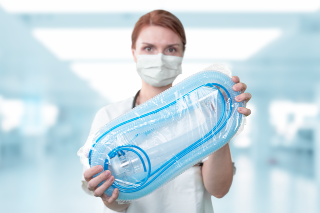
Preassembled. And ready to use

Our preassembled breathing circuit sets include the essential consumables to operate the ventilator, conveniently packaged in one single bag.

All our essential consumables are specially developed for Hamilton Medical ventilators with guaranteed manufacturer quality.

Automation; Hand turns knob button clockwise

Less knob-turning. More adaptations to your patient

To manage ventilation you usually have to set multiple parameters, such as pressure, volume, inspiratory and expiratory triggers, cuff pressure, and more. And each time your patient's condition changes, you have to make one or even several readjustments.

To simplify this process and reduce the knob-turning, we have created a range of solutions:

Adaptive Support Ventilation (ASV) is a ventilation mode that provides continuous adaptation of respiratory rate, tidal volume, and inspiratory time, depending on the patient’s lung mechanics and effort. ASV has been shown to shorten the duration of mechanical ventilation in various patient populations with fewer manual settings (​Kirakli C, Naz I, Ediboglu O, Tatar D, Budak A, Tellioglu E. A randomized controlled trial comparing the ventilation duration between adaptive support ventilation and pressure assist/control ventilation in medical patients in the ICU. Chest. 2015;147(6):1503-1509. doi:10.1378/chest.14-25991​, Tam MK, Wong WT, Gomersall CD, et al. A randomized controlled trial of 2 protocols for weaning cardiac surgical patients receiving adaptive support ventilation. J Crit Care. 2016;33:163-168. doi:10.1016/j.jcrc.2016.01.0182​, Zhu F, Gomersall CD, Ng SK, Underwood MJ, Lee A. A randomized controlled trial of adaptive support ventilation mode to wean patients after fast-track cardiac valvular surgery. Anesthesiology. 2015;122(4):832-840. doi:10.1097/ALN.00000000000005893​).

Conventional solutions for cuff pressure management require you to monitor and adjust cuff pressure by hand.

IntelliCuff secures your patient’s airway (Chenelle CT, Oto J, Sulemanji D, Fisher DF, Kacmarek RM. Evaluation of an automated endotracheal tube cuff controller during simulated mechanical ventilation. Respir Care. 2015;60(2):183-190. doi:10.4187/respcare.033874​) by continuously measuring and automatically maintaining the set cuff pressure for adult, pediatric, and neonatal patients.

Professional interacting with touch-screen

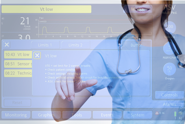
Help is near! On-screen troubleshooting

Whenever there is a problem, the ventilator alerts you using the alarm lamp, sound, and message bar.

The on-screen help offers you suggestions on how to resolve the alarm.

Patient in wheelchair with ventilator

Farewell ventilator! Tools to implement your weaning protocols

We want our ventilator to leave your patient’s side as quickly as possible. That is why we provide you with tools to help you implement your weaning protocol.

These include visual aids and ventilation modes designed to encourage spontaneous breathing.
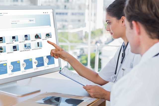
Professionals looking into Hamilton Medical e-learnings

Get the hang of it! Learning paths and educational content

Our online Academy offers easy-to-follow learning paths to familiarize you with Hamilton Medical products and technologies as quickly as possible.

For the future

Illustration of a compass pointing towards the future

Constant evolution. Expanding your ventilator’s capabilities

We are constantly working on further evolving our products. New features are added and existing features improved to ensure you always have access to the latest ventilation technology over your ventilator’s lifetime.
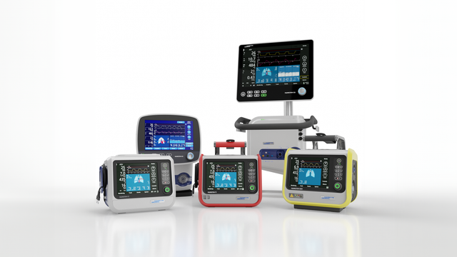
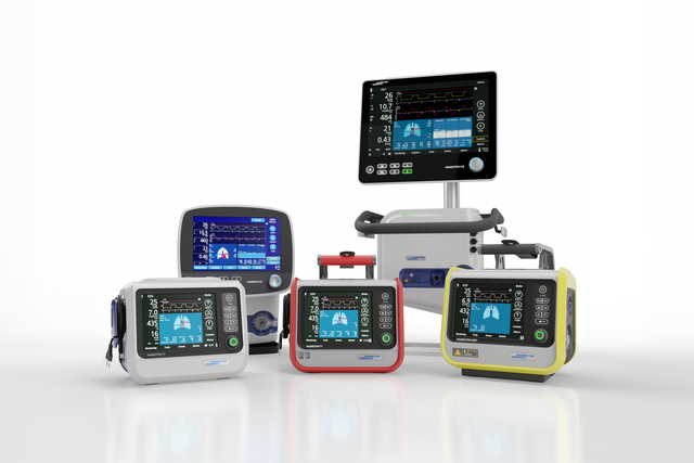
How we keep your ventilator up-to-date
Hamilton ventilation family Hamilton ventilation family

Know one, know them all. A universal user interface

Whether it is in the ICU, in the MRI suite, or during transport, the user interface of all Hamilton Medical ventilators works in the same way.

Our Ventilation Cockpit integrates complex data into intuitive visualizations.

For the complete solution

Fully integrated accessories

We develop our accessories for the highest possible patient safety and ease of use in mind. Whenever possible, we integrate them with our ventilators to simplify operation of the complete ventilator system.

Our consumables

All Hamilton Medical Originals are designed for optimal performance with Hamilton Medical ventilators. To ensure maximum user satisfaction and patient safety, we strive for the highest quality and safety standards.
Photo of an employee

Talk to our experts. Let's discuss your needs

Our team of Ventilation Geeks is happy to assist you in choosing the perfect ventilator for your clinical care setting and helping you meet your therapy goals. Get a personalized price quote or schedule a callback for more information.

A randomized controlled trial comparing the ventilation duration between adaptive support ventilation and pressure assist/control ventilation in medical patients in the ICU.

Kirakli C, Naz I, Ediboglu O, Tatar D, Budak A, Tellioglu E. A randomized controlled trial comparing the ventilation duration between adaptive support ventilation and pressure assist/control ventilation in medical patients in the ICU. Chest. 2015;147(6):1503-1509. doi:10.1378/chest.14-2599


BACKGROUND

Adaptive support ventilation (ASV) is a closed loop mode of mechanical ventilation (MV) that provides a target minute ventilation by automatically adapting inspiratory pressure and respiratory rate with the minimum work of breathing on the part of the patient. The aim of this study was to determine the effect of ASV on total MV duration when compared with pressure assist/control ventilation.

METHODS

Adult medical patients intubated and mechanically ventilated for > 24 h in a medical ICU were randomized to either ASV or pressure assist/control ventilation. Sedation and medical treatment were standardized for each group. Primary outcome was the total MV duration. Secondary outcomes were the weaning duration, number of manual settings of the ventilator, and weaning success rates.

RESULTS

Two hundred twenty-nine patients were included. Median MV duration until weaning, weaning duration, and total MV duration were significantly shorter in the ASV group (67 [43-94] h vs 92 [61-165] h, P = .003; 2 [2-2] h vs 2 [2-80] h, P = .001; and 4 [2-6] days vs 4 [3-9] days, P = .016, respectively). Patients in the ASV group required fewer total number of manual settings on the ventilator to reach the desired pH and Paco2 levels (2 [1-2] vs 3 [2-5], P < .001). The number of patients extubated successfully on the first attempt was significantly higher in the ASV group (P = .001). Weaning success and mortality at day 28 were comparable between the two groups.

CONCLUSIONS

In medical patients in the ICU, ASV may shorten the duration of weaning and total MV duration with a fewer number of manual ventilator settings.

TRIAL REGISTRY

ClinicalTrials.gov; No.: NCT01472302; URL: www.clinicaltrials.gov.

A randomized controlled trial of 2 protocols for weaning cardiac surgical patients receiving adaptive support ventilation.

Tam MK, Wong WT, Gomersall CD, et al. A randomized controlled trial of 2 protocols for weaning cardiac surgical patients receiving adaptive support ventilation. J Crit Care. 2016;33:163-168. doi:10.1016/j.jcrc.2016.01.018


PURPOSE

This study aims to compare the effectiveness of weaning with adaptive support ventilation (ASV) incorporating progressively reduced or constant target minute ventilation in the protocol in postoperative care after cardiac surgery.

MATERIAL AND METHODS

A randomized controlled unblinded study of 52 patients after elective coronary artery bypass surgery was carried out to determine whether a protocol incorporating a decremental target minute ventilation (DTMV) results in more rapid weaning of patients ventilated in ASV mode compared to a protocol incorporating a constant target minute ventilation.

RESULTS

Median duration of mechanical ventilation (145 vs 309 minutes; P = .001) and intubation (225 vs 423 minutes; P = .005) were significantly shorter in the DTMV group. There was no difference in adverse effects (42% vs 46%) or mortality (0% vs 0%) between the 2 groups.

CONCLUSIONS

Use of a DTMV protocol for postoperative ventilation of cardiac surgical patients in ASV mode results in a shorter duration of ventilation and intubation without evidence of increased risk of adverse effects.

A randomized controlled trial of adaptive support ventilation mode to wean patients after fast-track cardiac valvular surgery.

Zhu F, Gomersall CD, Ng SK, Underwood MJ, Lee A. A randomized controlled trial of adaptive support ventilation mode to wean patients after fast-track cardiac valvular surgery. Anesthesiology. 2015;122(4):832-840. doi:10.1097/ALN.0000000000000589


BACKGROUND

Adaptive support ventilation can speed weaning after coronary artery surgery compared with protocolized weaning using other modes. There are no data to support this mode of weaning after cardiac valvular surgery. Furthermore, control group weaning times have been long, suggesting that the results may reflect control group protocols that delay weaning rather than a real advantage of adaptive support ventilation.

METHODS

Randomized (computer-generated sequence and sealed opaque envelopes), parallel-arm, unblinded trial of adaptive support ventilation versus physician-directed weaning after adult fast-track cardiac valvular surgery. The primary outcome was duration of mechanical ventilation. Patients aged 18 to 80 yr without significant renal, liver, or lung disease or severe impairment of left ventricular function undergoing uncomplicated elective valve surgery were eligible. Care was standardized, except postoperative ventilation. In the adaptive support ventilation group, target minute ventilation and inspired oxygen concentration were adjusted according to blood gases. A spontaneous breathing trial was carried out when the total inspiratory pressure of 15 cm H2O or less with positive end-expiratory pressure of 5 cm H2O. In the control group, the duty physician made all ventilatory decisions.

RESULTS

Median duration of ventilation was statistically significantly shorter (P = 0.013) in the adaptive support ventilation group (205 [141 to 295] min, n = 30) than that in controls (342 [214 to 491] min, n = 31). Manual ventilator changes and alarms were less common in the adaptive support ventilation group, and arterial blood gas estimations were more common.

CONCLUSION

Adaptive support ventilation reduces ventilation time by more than 2 h in patients who have undergone fast-track cardiac valvular surgery while reducing the number of manual ventilator changes and alarms.

Evaluation of an automated endotracheal tube cuff controller during simulated mechanical ventilation.

Chenelle CT, Oto J, Sulemanji D, Fisher DF, Kacmarek RM. Evaluation of an automated endotracheal tube cuff controller during simulated mechanical ventilation. Respir Care. 2015;60(2):183-190. doi:10.4187/respcare.03387


BACKGROUND

Maintaining endotracheal tube cuff pressure within a narrow range is an important factor in patient care. The goal of this study was to evaluate the IntelliCuff against the manual technique for maintaining cuff pressure during simulated mechanical ventilation with and without movement.

METHODS

The IntelliCuff was compared to the manual technique of a manometer and syringe. Two independent studies were performed during mechanical ventilation: part 1, a 2-h trial incorporating continuous mannikin head movement; and part 2, an 8-h trial using a stationary trachea model. We set cuff pressure to 25 cm H2O, PEEP to 10 cm H2O, and peak inspiratory pressures to 20, 30, and 40 cm H2O. Clinical importance was defined as both statistically significant (P<.05) and clinically significant (pressure change [Δ]>10%).

RESULTS

In part 1, the change in cuff pressure from before to after ventilation was clinically important for the manual technique (P<.001, Δ=-39.6%) but not for the IntelliCuff (P=.02, Δ=3.5%). In part 2, the change in cuff pressure from before to after ventilation was clinically important for the manual technique (P=.004, Δ=-14.39%) but not for the IntelliCuff (P=.20, Δ=5.65%).

CONCLUSIONS

There was a clinically important drop in manually set cuff pressure during simulated mechanical ventilation in a stationary model and an even larger drop with movement, but this was significantly reduced by the IntelliCuff in both scenarios. Additionally, we observed that cuff pressure varied directly with inspiratory airway pressure for both techniques, leading to elevated average cuff pressures.

New frontiers in aerosol delivery during mechanical ventilation.

Dhand R. New frontiers in aerosol delivery during mechanical ventilation. Respir Care. 2004;49(6):666-677.

The scientific basis for inhalation therapy in mechanically-ventilated patients is now firmly established. A variety of new devices that deliver drugs to the lung with high efficiency could be employed for drug delivery during mechanical ventilation. Encapsulation of drugs within liposomes could increase the amount of drug delivered, prolong the effect of a dose, and minimize adverse effects. With improved inhalation devices and surfactant formulations, inhaled surfactant could be employed for several indications in mechanically-ventilated patients. Research is unraveling the causes of some disorders that have been poorly understood, and our improved understanding of the causal mechanisms of various respiratory disorders will provide new applications for inhaled therapies.

Advanced nebulizer designs employing vibrating mesh/aperture plate technologies for aerosol generation.

Waldrep JC, Dhand R. Advanced nebulizer designs employing vibrating mesh/aperture plate technologies for aerosol generation. Curr Drug Deliv. 2008;5(2):114-119. doi:10.2174/156720108783954815

Recent technological advances and improved nebulizer designs have overcome many limitations of jet nebulizers. Newer devices employ a vibrating mesh or aperture plate (VM/AP) for the generation of therapeutic aerosols with consistent, increased efficiency, predominant aerosol fine particle fractions, low residuals, and the ability to nebulize even microliter volumes. These enhancements are achieved through several different design features and include improvements that promote patient compliance, such as compact design, portability, shorter treatment durations, and quiet operation. Current VM/AP devices in clinical use are the Omron MicroAir, the Nektar Aeroneb, and the Pari eFlow. However, some devices are only approved for use with specific medications. Development of "smart nebulizers" such as the Respironics I-neb couple VM technologies with coordinated delivery and optimized inhalation patterns to enhance inhaled drug delivery of specialized, expensive formulations. Ongoing development of advanced aerosol technologies should improve clinical outcomes and continue to expand therapeutic options as newer inhaled drugs become available.