Hemos detectado que visita nuestro sitio web desde Estados Unidos.
Disponemos de una versión específica del sitio web para su país (Estados Unidos).

Cambiar a Estados Unidos
 Material fungible

Sensores de flujo. Medición de presión y flujo proximal

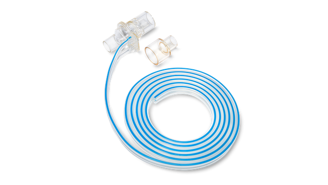
Sensor de flujo

¡Acérquese! Medición de flujo proximal

El sensor de flujo proximal ha sido la piedra angular de nuestros respiradores desde 1983. Todo el proceso de ventilación depende de la medición y precisión del sensor de flujo, que proporciona datos procedentes de la apertura de las vías aéreas.

La obtención de datos de presión, flujo y volumen precisos es fundamental para emitir un diagnóstico correcto y evitar los efectos secundarios habituales consecuencia de la configuración incorrecta del respirador. También admite algunas de nuestra tecnologías avanzadas, como los modos ASV e INTELLiVENT-ASV, además de IntelliSync+ y P/V Tool.

Sensor de flujo

La precisión es prioritaria. Sus pacientes dependen de ella.

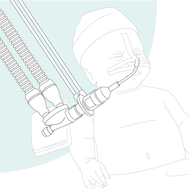
Nuestros respiradores miden el flujo y la presión cerca de la vía aérea del paciente. Los estudios han demostrado que los volúmenes tidales de los pacientes con ventilación se deben determinar con un sensor de flujo ubicado en el tubo endotraqueal (Cannon ML, Cornell J, Tripp-Hamel DS, et al. Tidal volumes for ventilated infants should be determined with a pneumotachometer placed at the endotracheal tube. Am J Respir Crit Care Med. 2000;162(6):2109-2112. doi:10.1164/ajrccm.162.6.99061121, Gammage, Gary W.; Banner, Michael J.; Blanch, Paul B.; Kirby, Robert R. VENTILATOR DISPLAYED TIDAL VOLUME—WHAT YOU SEE MAY NOT BE WHAT YOU GET, Critical Care Medicine: April 1988 - Volume 16 - Issue 4 - p 454 2)

Ilustración gráfica: mujer reflexionando sobre una pregunta

¿Tiene alguna prueba? Datos clínicos

La determinación precisa del volumen tidal espiratorio (VTE) es fundamental (Cannon ML, Cornell J, Tripp-Hamel DS, et al. Tidal volumes for ventilated infants should be determined with a pneumotachometer placed at the endotracheal tube. Am J Respir Crit Care Med. 2000;162(6):2109-2112. doi:10.1164/ajrccm.162.6.99061121), especialmente en situaciones donde se administran únicamente volúmenes tidales pequeños (lactantes, neonatos y pacientes con SDRA). Con los sensores de flujo de Hamilton Medical, puede medir el VTE cerca de la vía aérea del paciente para obtener un valor más preciso.

Beneficios para usted:

  • La colocación proximal elimina los efectos de la compliance del circuito respiratorio en las mediciones de flujo y volumen (Cannon ML, Cornell J, Tripp-Hamel DS, et al. Tidal volumes for ventilated infants should be determined with a pneumotachometer placed at the endotracheal tube. Am J Respir Crit Care Med. 2000;162(6):2109-2112. doi:10.1164/ajrccm.162.6.99061121)
  • La medición del VTE se ve sometida a una menor resistencia del sistema respiratorio (Nève V, Leclerc F, Noizet O, et al. Influence of respiratory system impedance on volume and pressure delivered at the Y piece in ventilated infants. Pediatric Critical Care Med. 2003;4(4):418-425. doi:10.1097/01.PCC.0000090289.98377.153)
  • Se producen menos fugas que puedan alterar el resultado (Al-Majed SI, Thompson JE, Watson KF, Randolph AG. Effect of lung compliance and endotracheal tube leakage on measurement of tidal volume. Crit Care. 2004;8(6):R398-R402. doi:10.1186/cc295444)

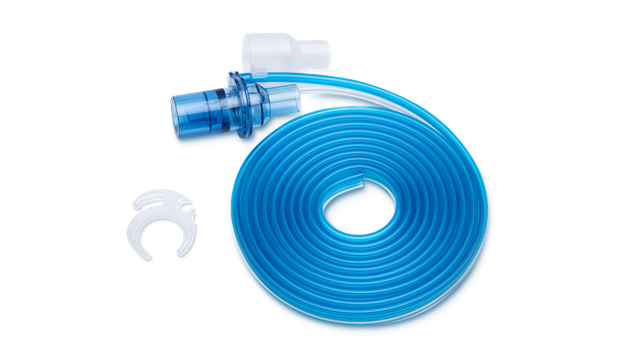
Nuestra gama de sensores de flujo

Ofrecemos material fungible de Hamilton Medical para pacientes adultos, pediátricos y neonatos. Puede elegir entre productos reutilizables o desechables, en función de las políticas de su centro sanitario.
Dr. Robert Lopez

Testimonios de clientes

Los sensores de flujo desechables de Hamilton Medical ayudan a evitar la contaminación cruzada, ya que no hay que preocuparse de que vuelvan a utilizarse en otro paciente.

Dr. Robert Lopez

Director del departamento de Asistencia Respiratoria hasta 2018
University Medical Center, Lubbock (TX), EE. UU.
Tidal volumes for ventilated infants should be determined with a pneumotachometer placed at the endotracheal tube.

Cannon ML, Cornell J, Tripp-Hamel DS, et al. Tidal volumes for ventilated infants should be determined with a pneumotachometer placed at the endotracheal tube. Am J Respir Crit Care Med. 2000;162(6):2109-2112. doi:10.1164/ajrccm.162.6.9906112

Many ventilators measure expired tidal volume (VT) without compensation either for the compliance of the ventilator circuit or for variations in the circuit setup. We hypothesized that the exhaled VT measured with a conventional ventilator at the expiratory valve would differ significantly from the exhaled VT measured with a pneumotachometer placed at the endotracheal tube. To investigate this we studied 98 infants and children requiring conventional ventilation. We used linear regression analysis to compare the VT obtained with the pneumotachometer with the ventilator-measured volume. An additional comparison was made between the pneumotachometer volume and a calculated effective VT. For infant circuits (n = 70), our analysis revealed a poor correlation between the expiratory VT measured with the pneumotachometer and the ventilator-measured volume (r(2) = 0.54). Similarly, the expiratory VT measured with the pneumotachometer did not correlate with the calculated effective volume (r(2) = 0.58). For pediatric circuits (n = 28), there was improved correlation between the expiratory VT measured with the pneumotachometer and both the ventilator-measured volume and the calculated effective VT (r(2) = 0.84 and r(2) = 0.85, respectively). The data demonstrate a significant discrepancy between expiratory VT measured at a ventilator and that measured with a pneumotachometer placed at the endotracheal tube in infants. Correcting for the compliance of the ventilator circuit by calculating the effective VT did not alter this discrepancy. In conventionally ventilated infants, exhaled VT should be determined with a pneumotachometer placed at the airway.

Ventilator displayed tidal volume: What you see may not be what you get.

Gammage, Gary W.; Banner, Michael J.; Blanch, Paul B.; Kirby, Robert R. VENTILATOR DISPLAYED TIDAL VOLUME—WHAT YOU SEE MAY NOT BE WHAT YOU GET, Critical Care Medicine: April 1988 - Volume 16 - Issue 4 - p 454

Influence of respiratory system impedance on volume and pressure delivered at the Y piece in ventilated infants.

Nève V, Leclerc F, Noizet O, et al. Influence of respiratory system impedance on volume and pressure delivered at the Y piece in ventilated infants. Pediatr Crit Care Med. 2003;4(4):418-425. doi:10.1097/01.PCC.0000090289.98377.15


OBJECTIVES

Tidal volume (VT) delivered to infants' airways are overestimated and pressure underestimated when measured in the ventilator and not at the Y piece. This study aimed at evaluating the influence of respiratory system impedance on expiratory VT (VTE) and pressure measurement difference.

DESIGN

Prospective observational study.

SETTING

Pediatric intensive care unit at a university hospital.

PATIENTS

Data were collected between February 2000 and October 2001 for 30 infants (range, 1-23 months) ventilated in the pressure-controlled or volume-controlled mode.

INTERVENTIONS

Measurements of VTE, pressure obtained at the same time at the Y piece and on the ventilator Servo 300, were collected in ventilated infants. Respiratory system impedance was calculated from data obtained at the Y piece. Circuit compliance was measured in vitro. VTEs were corrected for compressible volume.

MEASUREMENTS AND RESULTS

VTEs were overestimated by the Servo 300 in the pressure-controlled and volume-controlled modes (from 5% to 62% of the value displayed on Servo 300). Maximal inspiratory pressures were underestimated by the Servo 300 in the pressure-controlled mode (difference from -2 to +19 cm H(2)O). Measurement difference increased with increasing respiratory system impedance. Ventilator VTE corrected for circuit compliance did not offer a sufficiently accurate estimation of VTE at the Y piece.

CONCLUSIONS

VT and pressure measurements must be performed at the Y piece, especially in infants with increased respiratory system impedance (i.e., decreased respiratory system compliance or increased resistance). Correcting VTE for circuit compliance cannot replace measurement of VT at the Y piece.

Effect of lung compliance and endotracheal tube leakage on measurement of tidal volume.

Al-Majed SI, Thompson JE, Watson KF, Randolph AG. Effect of lung compliance and endotracheal tube leakage on measurement of tidal volume. Crit Care. 2004;8(6):R398-R402. doi:10.1186/cc2954


INTRODUCTION

The objective of this laboratory study was to measure the effect of decreased lung compliance and endotracheal tube (ETT) leakage on measured exhaled tidal volume at the airway and at the ventilator, in a research study with a test lung.

METHODS

The subjects were infant, adult and pediatric test lungs. In the test lung model, lung compliances were set to normal and to levels seen in acute respiratory distress syndrome. Set tidal volume was 6 ml/kg across a range of simulated weights and ETT sizes. Data were recorded from both the ventilator light-emitting diode display and the CO2SMO Plus monitor display by a single observer. Effective tidal volume was calculated from a standard equation.

RESULTS

In all test lung models, exhaled tidal volume measured at the airway decreased markedly with decreasing lung compliance, but measurement at the ventilator showed minimal change. In the absence of a simulated ETT leak, calculation of the effective tidal volume led to measurements very similar to exhaled tidal volume measured at the ETT. With a simulated ETT tube leak, the effective tidal volume markedly overestimated tidal volume measured at the airway.

CONCLUSION

Previous investigators have emphasized the need to measure tidal volume at the ETT for all children. When ETT leakage is minimal, it seems from our simulated lung models that calculation of effective tidal volume would give similar readings to tidal volume measured at the airway, even in small patients. Future studies of tidal volume measurement accuracy in mechanically ventilated children should control for the degree of ETT leakage.