Nous avons remarqué que vous aviez consulté notre site internet depuis le pays États-Unis.
Nous proposons une version du site internet propre à votre pays (États-Unis).

Passer à États-Unis
 Technologies

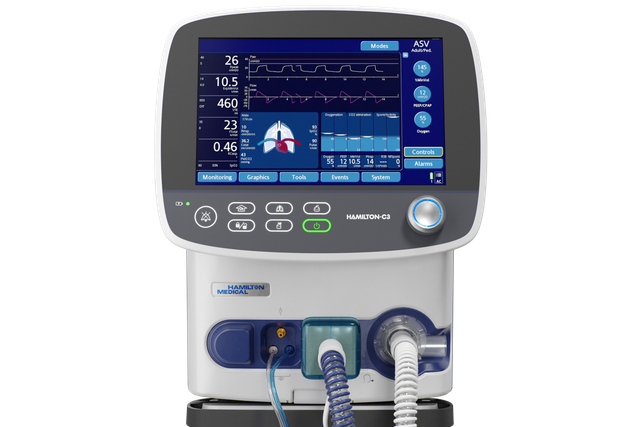
Le Ventilation Cockpit. Afficher clairement les données complexes

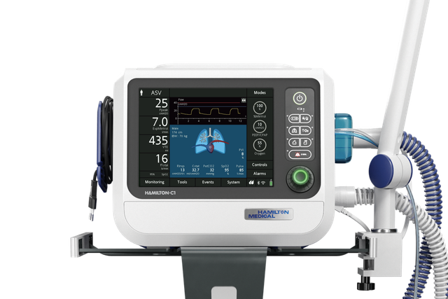
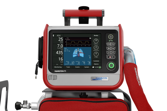
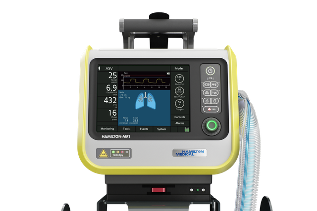
Famille de ventilateurs Hamilton

Notre mission. Une interface pour tous les ventilateurs

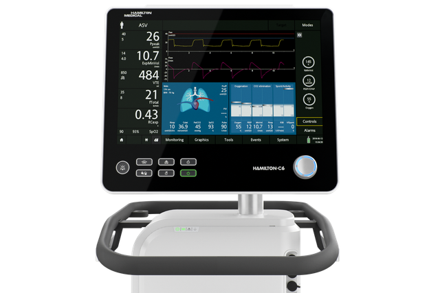
L'interface utilisateur fonctionne de la même manière sur tous nos ventilateurs et ce, indépendamment de son utilisation en USI, en salle d'examen IRM ou lors de déplacements.

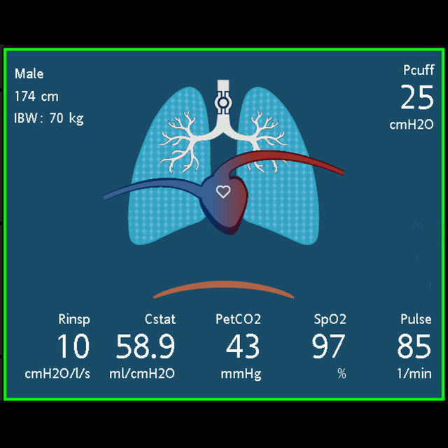
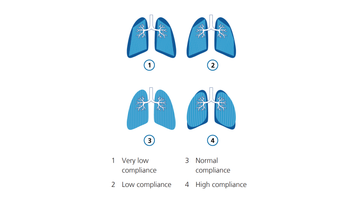
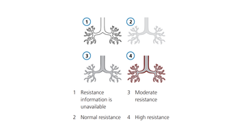
Notre Ventilation Cockpit intègre des données complexes qui sont représentées visuellement de façon intuitive.

L'inspiration. Représentation graphique de données complexes

Une étude a montré que les affichages de chiffres et de formes d'ondes uniquement ne sont pas suffisants pour fournir une aide optimale aux médecins (Drews FA, Westenskow DR. The right picture is worth a thousand numbers: data displays in anesthesia. Hum Factors. 2006;48(1):59-71. doi:10.1518/0018720067764122701​). Une autre solution consiste à afficher des graphiques qui représentent visuellement des données, ce qui aide les médecins à détecter et à traiter plus rapidement des événements respiratoires indésirables, tout en diminuant sensiblement la charge de travail (Wachter SB, Johnson K, Albert R, Syroid N, Drews F, Westenskow D. The evaluation of a pulmonary display to detect adverse respiratory events using high resolution human simulator. J Am Med Inform Assoc. 2006;13(6):635-642. doi:10.1197/jamia.M21232​).

Notre Ventilation Cockpit s'inspire du cockpit des avions dans lesquels des données complexes sont intégrées et représentées visuellement de façon simplifiée.
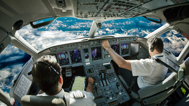
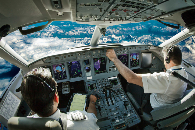
Aperçu du cockpit d'un avion avec des pilotes utilisant le cockpit. Aperçu du cockpit d'un avion avec des pilotes utilisant le cockpit.
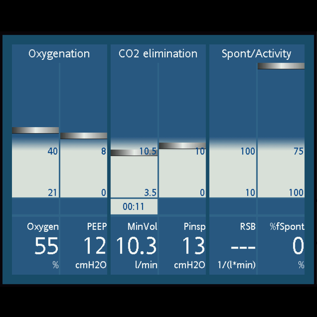
Panneau État Vent

Prêt pour le sevrage ? Le panneau État Vent

Le panneau État Vent affiche six paramètres relatifs à la dépendance du patient au ventilateur, notamment l’oxygénation, l’élimination du CO2 et l’activité du patient.

Une réglette variable à l'intérieur de la colonne représente la valeur d'un paramètre donné à chaque cycle. Lorsque cette réglette pénètre dans la zone de sevrage grise, un chronomètre démarre et indique le temps écoulé dans la zone de sevrage pour ce paramètre.

Lorsque toutes les valeurs se trouvent dans la zone de sevrage, le cadre entourant le panneau devient vert, indiquant que des épreuves de ventilation spontanée peuvent être envisagées.

Craig Jolly

Témoignages de clients

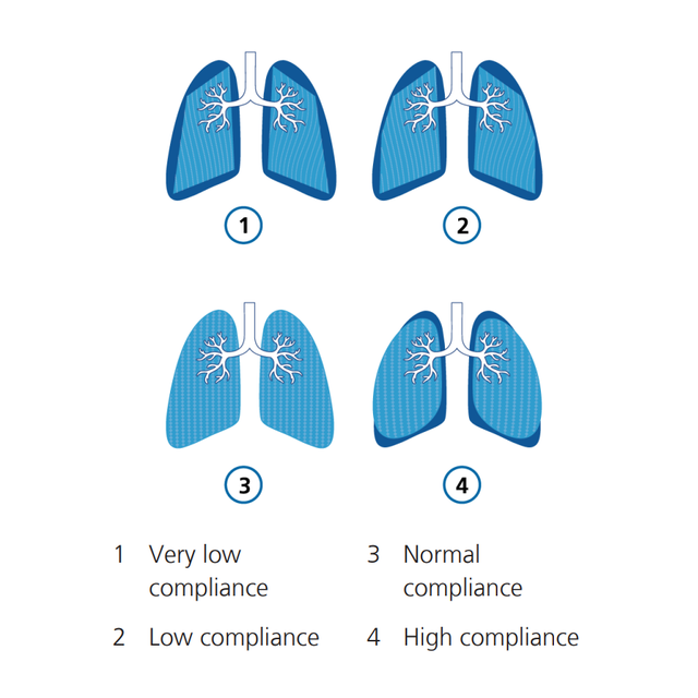
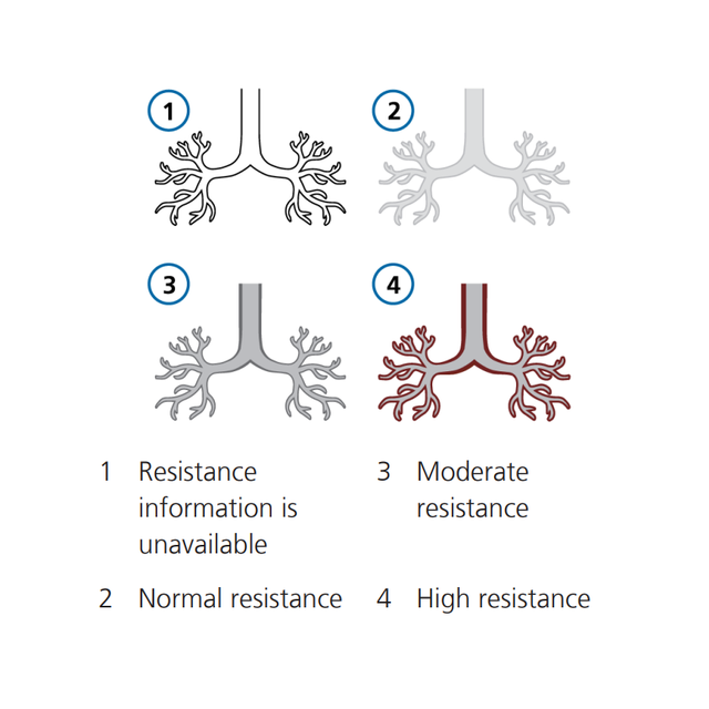
D'après mon expérience, la fonction Poumon dynamique est très utile car nous ne sommes pas tous toujours en mesure d'interpréter les chiffres, en particulier les thérapeutes inexpérimentés. Mais ils peuvent comprendre les images.

Craig Jolly

Coordinateur de la formation clinique
University Medical Center, Lubbock (Texas), États-Unis

Disponibilité

Le Ventilation Cockpit est une fonction standard équipant tous nos ventilateurs de soins intensifs.

The right picture is worth a thousand numbers: data displays in anesthesia.

Drews FA, Westenskow DR. The right picture is worth a thousand numbers: data displays in anesthesia. Hum Factors. 2006;48(1):59-71. doi:10.1518/001872006776412270


OBJECTIVE

To review the literature on data displays in anesthesia identifying issues and developing design recommendations.

BACKGROUND

Unexpected incidents are common in critical care medicine. Adverse outcomes are frequently the catastrophic endpoints of an "evolving" chain of subtle incidents. One strategy to reduce the likelihood of an adverse patient outcome during anesthesia is to improve the anesthesiologist's ability to detect, diagnose, and treat critical incidents.

METHOD

A literature review and analysis of data displays.

RESULTS

Current numerical and waveform displays do not support anesthesiologists optimally. An alternative is graphical displays that functionally integrate variables into objects. In a well-designed graphic object, deviations from normal are shown by distortions in the object's symmetry. The emerging patterns that result from distorted symmetry facilitate the correct diagnosis. When treatment is effective, an object's shape is restored to normal. Graphical displays can be an effective tool in supporting anesthesiologists' situation awareness.

CONCLUSION

Problems related to graphical displays have delayed their use in anesthesia, including the lack of conclusive clinical evidence of their value. However, currently more evidence is accumulating that graphical displays have the potential to improve clinical performance. The successful development of these graphical displays takes into account task requirements, a user's perceptual processes, and task-specific cognition.

APPLICATION

This paper provides suggestions for the development of more effective displays in anesthesiology. Graphical displays can increase the anesthesiologist's situation awareness and improve clinical performance. Clinical use of these displays has the potential to significantly improve patient safety.

The evaluation of a pulmonary display to detect adverse respiratory events using high resolution human simulator.

Wachter SB, Johnson K, Albert R, Syroid N, Drews F, Westenskow D. The evaluation of a pulmonary display to detect adverse respiratory events using high resolution human simulator. J Am Med Inform Assoc. 2006;13(6):635-642. doi:10.1197/jamia.M2123


OBJECTIVE

Authors developed a picture-graphics display for pulmonary function to present typical respiratory data used in perioperative and intensive care environments. The display utilizes color, shape and emergent alerting to highlight abnormal pulmonary physiology. The display serves as an adjunct to traditional operating room displays and monitors.

DESIGN

To evaluate the prototype, nineteen clinician volunteers each managed four adverse respiratory events and one normal event using a high-resolution patient simulator which included the new displays (intervention subjects) and traditional displays (control subjects). Between-group comparisons included (i) time to diagnosis and treatment for each adverse respiratory event; (ii) the number of unnecessary treatments during the normal scenario; and (iii) self-reported workload estimates while managing study events.

MEASUREMENTS

Two expert anesthesiologists reviewed video-taped transcriptions of the volunteers to determine time to treat and time to diagnosis. Time values were then compared between groups using a Mann-Whitney-U Test. Estimated workload for both groups was assessed using the NASA-TLX and compared between groups using an ANOVA. P-values < 0.05 were considered significant.

RESULTS

Clinician volunteers detected and treated obstructed endotracheal tubes and intrinsic PEEP problems faster with graphical rather than conventional displays (p < 0.05). During the normal scenario simulation, 3 clinicians using the graphical display, and 5 clinicians using the conventional display gave unnecessary treatments. Clinician-volunteers reported significantly lower subjective workloads using the graphical display for the obstructed endotracheal tube scenario (p < 0.001) and the intrinsic PEEP scenario (p < 0.03).

CONCLUSION

Authors conclude that the graphical pulmonary display may serve as a useful adjunct to traditional displays in identifying adverse respiratory events.