State accedendo al sito dal Paese Stati Uniti.
Per il vostro Paese (Stati Uniti) è disponibile anche una versione dedicata del sito.

Passare a Stati Uniti
 Tecnologie

Il Ventilation Cockpit: visualizzazione intuitiva di dati complessi

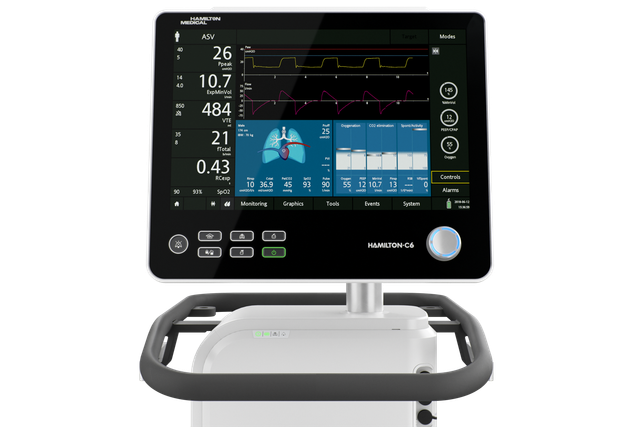
La famiglia di ventilatori Hamilton

La nostra visione: un'unica interfaccia per usarli tutti

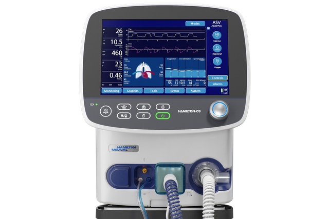
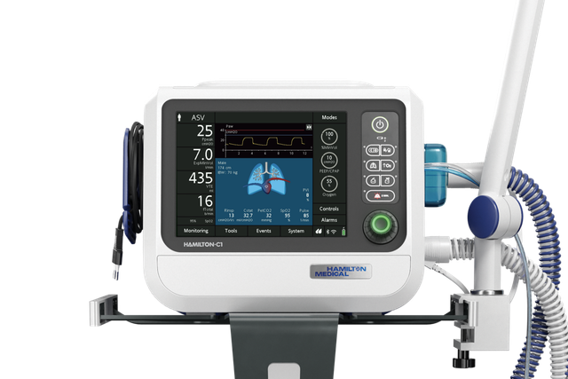
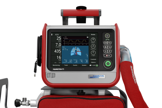
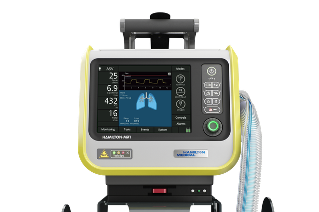
Che si utilizzi il dispositivo in terapia intensiva, nella sala per imaging RM o durante il trasporto, l'interfaccia di tutti i nostri ventilatori funziona allo stesso modo.

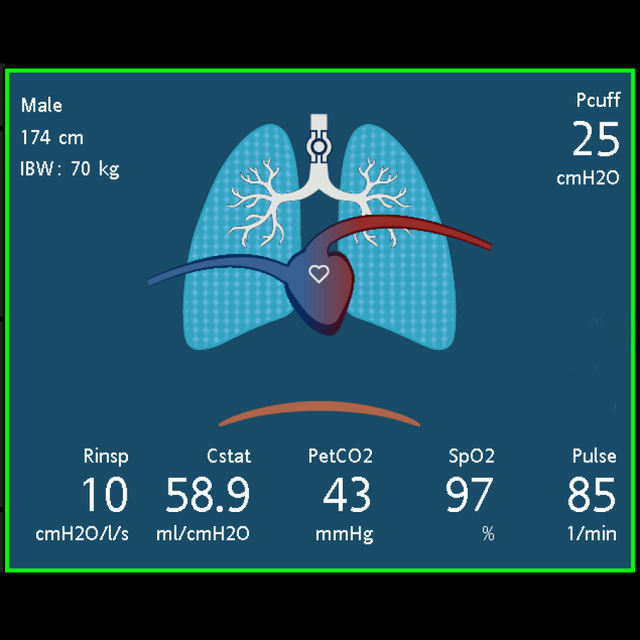
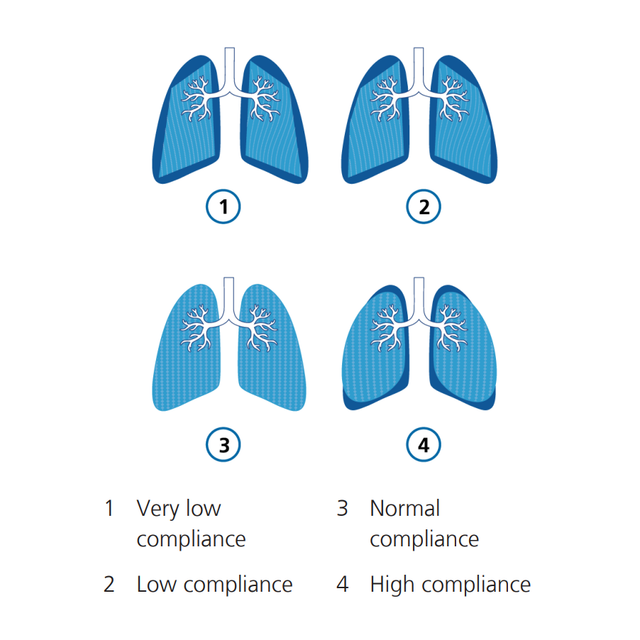
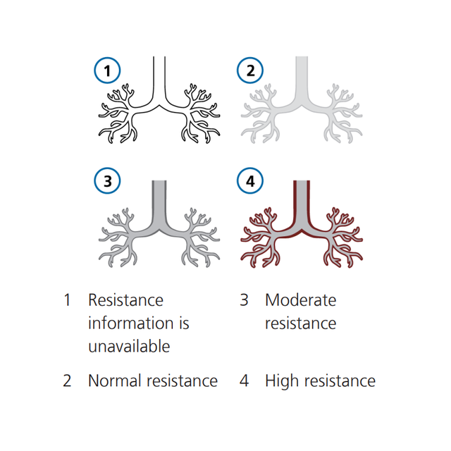
Il nostro Ventilation Cockpit visualizza dati complessi all'interno di viste intuitive.

L'ispirazione: rappresentare graficamente i dati più complessi

Uno studio ha rilevato che la sola visualizzazione dei valori numerici e delle curve non basta per sostenere in modo ottimale il lavoro dei medici (Drews FA, Westenskow DR. The right picture is worth a thousand numbers: data displays in anesthesia. Hum Factors. 2006;48(1):59-71. doi:10.1518/0018720067764122701). Come soluzione alternativa, le visualizzazioni grafiche che integrano i dati in un'immagine possono aiutare i medici a rilevare e trattare gli eventi respiratori avversi più rapidamente, con una riduzione del carico di lavoro percepito (Wachter SB, Johnson K, Albert R, Syroid N, Drews F, Westenskow D. The evaluation of a pulmonary display to detect adverse respiratory events using high resolution human simulator. J Am Med Inform Assoc. 2006;13(6):635-642. doi:10.1197/jamia.M21232).

Per il nostro Ventilation Cockpit abbiamo preso ispirazione dalle cabine di pilotaggio dei velivoli, che integrano dati complessi e li visualizzano in layout semplificati.
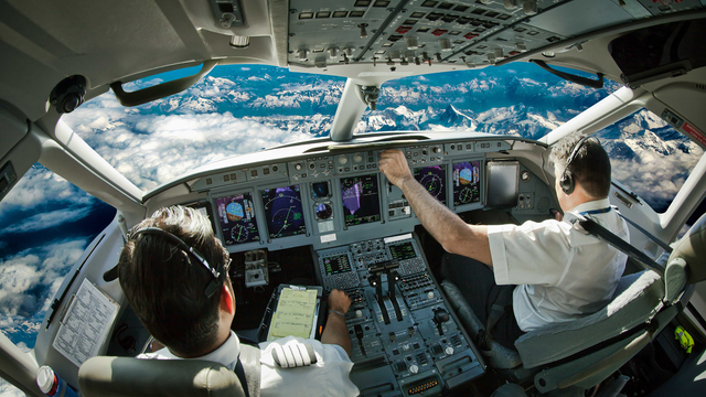
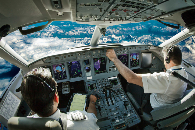
Foto della cabina di pilotaggio di un aereo, con i piloti che azionano i comandi del cockpit. Foto della cabina di pilotaggio di un aereo, con i piloti che azionano i comandi del cockpit.
Pannello StatoVent

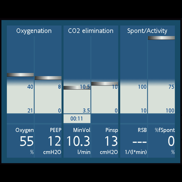
Tutti pronti per lo svezzamento? Il pannello StatoVent

Il pannello StatoVent visualizza sei parametri relativi alla dipendenza del paziente dal ventilatore, tra cui l'ossigenazione, l'eliminazione della CO2 e l'attività del paziente.

Un cursore fluttuante si sposta all'interno della colonna indicando il valore di un dato parametro respiro per respiro. Quando il cursore si trova nella zona grigia di svezzamento, si attiva un timer che indica da quanto tempo quel parametro si trova nella zona di svezzamento.

Quando tutti i valori rientrano nella zona di svezzamento, il pannello è circondato da un riquadro verde, per indicare la possibilità di condurre prove di respiro spontaneo o un'estubazione.

Craig Jolly

Cosa dicono i clienti

So per esperienza che il pannello PolmDin è davvero utile, perché non tutti riescono a interpretare sempre correttamente i valori numerici, soprattutto i terapisti alle prime armi. Ma comprendono al volo l'immagine.

Craig Jolly

Coordinatore formazione clinica
Centro medico universitario, Lubbock, Texas, USA

Disponibilità

Il Ventilation Cockpit è una funzione di serie su tutti i nostri ventilatori per la terapia intensiva.

The right picture is worth a thousand numbers: data displays in anesthesia.

Drews FA, Westenskow DR. The right picture is worth a thousand numbers: data displays in anesthesia. Hum Factors. 2006;48(1):59-71. doi:10.1518/001872006776412270


OBJECTIVE

To review the literature on data displays in anesthesia identifying issues and developing design recommendations.

BACKGROUND

Unexpected incidents are common in critical care medicine. Adverse outcomes are frequently the catastrophic endpoints of an "evolving" chain of subtle incidents. One strategy to reduce the likelihood of an adverse patient outcome during anesthesia is to improve the anesthesiologist's ability to detect, diagnose, and treat critical incidents.

METHOD

A literature review and analysis of data displays.

RESULTS

Current numerical and waveform displays do not support anesthesiologists optimally. An alternative is graphical displays that functionally integrate variables into objects. In a well-designed graphic object, deviations from normal are shown by distortions in the object's symmetry. The emerging patterns that result from distorted symmetry facilitate the correct diagnosis. When treatment is effective, an object's shape is restored to normal. Graphical displays can be an effective tool in supporting anesthesiologists' situation awareness.

CONCLUSION

Problems related to graphical displays have delayed their use in anesthesia, including the lack of conclusive clinical evidence of their value. However, currently more evidence is accumulating that graphical displays have the potential to improve clinical performance. The successful development of these graphical displays takes into account task requirements, a user's perceptual processes, and task-specific cognition.

APPLICATION

This paper provides suggestions for the development of more effective displays in anesthesiology. Graphical displays can increase the anesthesiologist's situation awareness and improve clinical performance. Clinical use of these displays has the potential to significantly improve patient safety.

The evaluation of a pulmonary display to detect adverse respiratory events using high resolution human simulator.

Wachter SB, Johnson K, Albert R, Syroid N, Drews F, Westenskow D. The evaluation of a pulmonary display to detect adverse respiratory events using high resolution human simulator. J Am Med Inform Assoc. 2006;13(6):635-642. doi:10.1197/jamia.M2123


OBJECTIVE

Authors developed a picture-graphics display for pulmonary function to present typical respiratory data used in perioperative and intensive care environments. The display utilizes color, shape and emergent alerting to highlight abnormal pulmonary physiology. The display serves as an adjunct to traditional operating room displays and monitors.

DESIGN

To evaluate the prototype, nineteen clinician volunteers each managed four adverse respiratory events and one normal event using a high-resolution patient simulator which included the new displays (intervention subjects) and traditional displays (control subjects). Between-group comparisons included (i) time to diagnosis and treatment for each adverse respiratory event; (ii) the number of unnecessary treatments during the normal scenario; and (iii) self-reported workload estimates while managing study events.

MEASUREMENTS

Two expert anesthesiologists reviewed video-taped transcriptions of the volunteers to determine time to treat and time to diagnosis. Time values were then compared between groups using a Mann-Whitney-U Test. Estimated workload for both groups was assessed using the NASA-TLX and compared between groups using an ANOVA. P-values < 0.05 were considered significant.

RESULTS

Clinician volunteers detected and treated obstructed endotracheal tubes and intrinsic PEEP problems faster with graphical rather than conventional displays (p < 0.05). During the normal scenario simulation, 3 clinicians using the graphical display, and 5 clinicians using the conventional display gave unnecessary treatments. Clinician-volunteers reported significantly lower subjective workloads using the graphical display for the obstructed endotracheal tube scenario (p < 0.001) and the intrinsic PEEP scenario (p < 0.03).

CONCLUSION

Authors conclude that the graphical pulmonary display may serve as a useful adjunct to traditional displays in identifying adverse respiratory events.