Indietro

Il metodo delle curve per la rilevazione dell'attività del paziente: è efficiente quanto la Pes?

Articolo

Autore: Caroline Brown, Giorgio Iotti

Data: 08.07.2022

L'asincronia tra paziente e ventilatore è un'eventualità comune nei pazienti ventilati meccanicamente (1, 2).

Il metodo delle curve per la rilevazione dell'attività del paziente: è efficiente quanto la Pes?

Contenuti chiave

  • L'idea di analizzare le curve di pressione e flusso per rilevare gli sforzi respiratori è stata formulata per la prima volta decenni fa, ma le evidenze sull'affidabilità di questo approccio emerse successivamente non sono determinanti.
  • In uno studio recente alcuni sperimentatori, utilizzando come riferimento la curva della Pes, hanno valutato un metodo sistematico basato sull'analisi delle curve utilizzabile al posto letto per la valutazione dell'attività del paziente e dell'interazione paziente‑ventilatore.
  • Il metodo delle curve ha consentito ai medici di rilevare una percentuale molto alta degli sforzi spontanei e si è dimostrato un mezzo altamente riproducibile e affidabile per identificare anche le asincronie lievi.

Una parte importante del trattamento

La mancata corrispondenza tra i tempi inspiratori ed espiratori di paziente e ventilatore può presentarsi sotto diverse forme, per esempio come ciclaggio ritardato, trigger automatico, doppio trigger o sforzo inefficace, ed è stato evidenziato che questa situazione ha un impatto sugli esiti dei pazienti (de Wit M, Miller KB, Green DA, Ostman HE, Gennings C, Epstein SK. Ineffective triggering predicts increased duration of mechanical ventilation. Crit Care Med. 2009;37(10):2740‑2745. doi:10.1097/ccm.0b013e3181a98a053, Blanch L, Villagra A, Sales B, et al. Asynchronies during mechanical ventilation are associated with mortality. Intensive Care Med. 2015;41(4):633‑641. doi:10.1007/s00134‑015‑3692‑64). Una parte importante del trattamento è quindi la possibilità di riconoscere queste asincronie e di regolare le impostazioni del ventilatore di conseguenza per migliorare l'interazione paziente‑ventilatore.

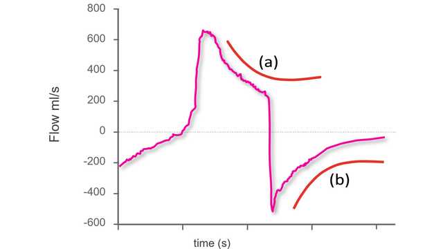
L'ipotesi di analizzare le curve di flusso e pressione delle vie aeree per rilevare gli sforzi espiratori e i relativi tempi è stata avanzata quasi trent'anni fa (Fabry B, Guttmann J, Eberhard L, Bauer T, Haberthür C, Wolff G. An analysis of desynchronization between the spontaneously breathing patient and ventilator during inspiratory pressure support. Chest. 1995;107(5):1387‑1394. doi:10.1378/chest.107.5.13875, Giannouli E, Webster K, Roberts D, Younes M. Response of ventilator‑dependent patients to different levels of pressure support and proportional assist. Am J Respir Crit Care Med. 1999;159(6):1716‑1725. doi:10.1164/ajrccm.159.6.97040256), ma le evidenze successive sull'affidabilità di questo approccio non sono determinanti (Thille AW, Rodriguez P, Cabello B, Lellouche F, Brochard L. Patient‑ventilator asynchrony during assisted mechanical ventilation. Intensive Care Med. 2006;32(10):1515‑1522. doi:10.1007/s00134‑006‑0301‑82​, Colombo D, Cammarota G, Alemani M, et al. Efficacy of ventilator waveforms observation in detecting patient‑ventilator asynchrony. Crit Care Med. 2011;39(11):2452‑2457. doi:10.1097/CCM.0b013e318225753c7). Secondo l'opinione più diffusa, è necessaria la misurazione della pressione esofagea; questo monitoraggio richiede però un'attrezzatura specifica e non è una normale prassi in ambito clinico. Uno studio recente condotto su 16 pazienti ha valutato se l'analisi delle curve fosse un mezzo riproducibile e affidabile per la rilevazione al posto letto dell'attività dei muscoli respiratori di un paziente (Mojoli F, Pozzi M, Orlando A, et al. Timing of inspiratory muscle activity detected from airway pressure and flow during pressure support ventilation: the waveform method. Crit Care. 2022;26(1):32. Pubblicato il 30 gennaio 2022. doi:10.1186/s13054‑022‑03895‑48).

Applicazione del metodo delle curve

Un elemento chiave di questa sperimentazione è stato l'utilizzo di un metodo sistematico per l'analisi delle curve di flusso e pressione delle vie aeree, costituito da cinque principi generali e una serie di regole specifiche definite in anticipo (che chiameremo "il metodo delle curve"). Tutti i pazienti erano ventilati in modalità a supporto di pressione con un catetere esofageo posizionato. Il metodo è stato applicato alle curve del flusso e della pressione delle vie aeree ottenute tramite un sensore prossimale; la pressione esofagea (Pes) è stata utilizzata come punto di riferimento. Per ogni paziente, tre ricercatori presi da un gruppo di quattro (tre medici senior e uno specializzando) hanno analizzato solo le curve di flusso e pressione, mentre il quarto ricercatore ha analizzato sia le curve di flusso e pressione sia la curva della Pes. I respiri sono stati classificati come normalmente assistiti, a trigger automatico, a doppio trigger o sforzi inefficaci. Nel caso dei respiri normalmente assistiti, sono state valutate anche le asincronie lievi (ritardo del trigger, ciclaggio prematuro e ciclaggio ritardato).

Endpoint e risultati

L'endpoint primario era la percentuale di sforzi spontanei rilevati utilizzando il metodo delle curve. Tra gli endpoint secondari sono stati inclusi l'accordo tra il metodo delle curve e il metodo di riferimento per quanto riguarda la rilevazione di sincronie maggiori e minori, oltre all'accordo reciproco delle valutazioni eseguite con il metodo delle curve da diversi valutatori.

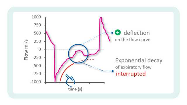
Sono stati registrati in totale 4.426 respiri. Utilizzando le misure di riferimento derivate dalla Pes, il 77,8% dei respiri è stato identificato come respiro correttamente rilevato dal ventilatore, il 22,1% come sforzo inefficace e lo 0,1% come respiro a trigger automatico. Con il metodo delle curve è stato possibile rilevare il 99,5% degli sforzi spontanei e tutti i respiri a trigger automatico tranne uno. L'accordo tra il metodo di riferimento e il metodo delle curve è stato molto elevato anche per la classificazione dei respiri come assistiti, a trigger automatico, a doppio trigger o sforzi inefficaci. L'indice di asincronia, calcolato come la somma dei respiri a trigger automatico, a doppio trigger e inefficaci divisa per il numero totale di respiri, è risultato pari a 5,9% e il valore ottenuto è stato il medesimo con il metodo delle curve e con quello della pressione esofagea. Il tempo di asincronia totale, calcolato come il tempo durante il quale paziente e ventilatore non erano sincronizzati diviso per il tempo di registrazione complessivo, era del 22,4%, con una componente del 92,1% dovuta alle asincronie lievi. Anche l'accordo tra i diversi operatori nella classificazione dei respiri è stato molto elevato.

In oltre il 90% dei casi, il metodo delle curve ha consentito ai ricercatori di identificare l'inizio e la fine degli sforzi respiratori con una precisione sufficiente a classificare correttamente anche le asincronie lievi (trigger ritardato, ciclaggio prematuro e ciclaggio ritardato).

Cosa ci dicono questi risultati?

Questo studio ha ottenuto alcuni importanti risultati Gli sperimentatori hanno evidenziato che il metodo delle curve consente ai medici di rilevare una percentuale molto alta degli sforzi respiratori spontanei e di valutare con precisione i tempi dell'attività del paziente. Anche nel caso di asincronie lievi, il metodo delle curve risulta altamente affidabile e riproducibile. L'importanza di questo risultato è sottolineata da un ulteriore dato rilevato dallo studio: la maggior parte del tempo di asincronia in modalità PSV era dovuto ad asincronie minori.

Non solo questi risultati dimostrano la riproducibilità del metodo delle curve (con un'elevato accordo inter‑operatore) ma evidenziano anche che una formazione nell'analisi delle curve secondo un metodo predefinito e sistematico gioca un ruolo fondamentale. Le evidenze hanno dimostrato che l'esperienza clinica nel trattamento dei pazienti ventilati meccanicamente non equivale necessariamente alla capacità di riconoscere le asincronie, che è complessivamente poco comune nei medici di terapia intensiva (Ramirez II, Arellano DH, Adasme RS, et al. Ability of ICU Health-Care Professionals to Identify Patient-Ventilator Asynchrony Using Waveform Analysis. Respir Care. 2017;62(2):144-149. doi:10.4187/respcare.047509). Nel momento in cui è stato condotto lo studio, uno degli operatori era semplicemente uno specializzando ma tutti gli operatori avevano almeno due anni di esperienza nell'analisi delle curve e utilizzavano un metodo sistematico con regole ben definite. Secondo gli autori questo è uno dei possibili motivi alla base delle differenze tra i risultati di questo studio e quelli ottenuti da Colombo et al. (Colombo D, Cammarota G, Alemani M, et al. Efficacy of ventilator waveforms observation in detecting patient-ventilator asynchrony. Crit Care Med. 2011;39(11):2452-2457. doi:10.1097/CCM.0b013e318225753c7), che hanno evidenziato un'elevata specificità ma una bassa sensibilità della rilevazione delle asincronie maggiori tramite l'osservazione delle curve.

Gli autori concludono che le curve di flusso d'aria e pressione delle vie aeree misurate a livello prossimale contengono informazioni sufficienti per valutare in modo accurato l'attività del paziente e l'interazione paziente‑ventilatore, ipotizzando che si adotti un metodo di analisi sistematico adeguato, come il "metodo delle curve".

Analisi continua con IntelliSync+

La tecnologia IntelliSync®+ integrata nei ventilatori Hamilton Medical ( IntelliSync+ è disponibile come funzione opzionale sui ventilatori meccanici HAMILTON‑C6 e HAMILTON‑G5, è invece una funzione standard sul ventilatore HAMILTON‑S1.A) analizza in modo continuativo il flusso e la pressione delle vie aeree a livello prossimale basandosi su principi analoghi a quelli utilizzati dal "metodo delle curve". È quindi in grado di identificare i segni precoci di uno sforzo inspiratorio o di un rilassamento del paziente e può pertanto iniziare di conseguenza l'inspirazione e il ciclaggio per passare all'espirazione. La funzione può essere attivata per l'automazione del solo trigger inspiratorio o espiratorio, oppure di entrambi.


Citazioni complete a fondo pagina: (Chao DC, Scheinhorn DJ, Stearn‑Hassenpflug M. Patient‑ventilator trigger asynchrony in prolonged mechanical ventilation. Chest. 1997;112(6):1592‑1599. doi:10.1378/chest.112.6.15921).

Scheda di riferimento sulle asincronie

Imparare a notare le più comuni asincronie: scheda di riferimento gratuita

La nostra scheda di riferimento sulle asincronie fornisce una panoramica sui tipi più comuni di asincronie, sulle loro cause e su come individuarle.

Note

  • A. IntelliSync+ è disponibile come funzione opzionale sui ventilatori meccanici HAMILTON‑C6 e HAMILTON‑G5, è invece una funzione standard sul ventilatore HAMILTON‑S1.

Bibliografia

  1. 1. Chao DC, Scheinhorn DJ, Stearn‑Hassenpflug M. Patient‑ventilator trigger asynchrony in prolonged mechanical ventilation. Chest. 1997;112(6):1592‑1599. doi:10.1378/chest.112.6.1592
  2. 2. Thille AW, Rodriguez P, Cabello B, Lellouche F, Brochard L. Patient‑ventilator asynchrony during assisted mechanical ventilation. Intensive Care Med. 2006;32(10):1515‑1522. doi:10.1007/s00134‑006‑0301‑8
  3. 3. de Wit M, Miller KB, Green DA, Ostman HE, Gennings C, Epstein SK. Ineffective triggering predicts increased duration of mechanical ventilation. Crit Care Med. 2009;37(10):2740‑2745. doi:10.1097/ccm.0b013e3181a98a05
  4. 4. Blanch L, Villagra A, Sales B, et al. Asynchronies during mechanical ventilation are associated with mortality. Intensive Care Med. 2015;41(4):633‑641. doi:10.1007/s00134‑015‑3692‑6
  5. 5. Fabry B, Guttmann J, Eberhard L, Bauer T, Haberthür C, Wolff G. An analysis of desynchronization between the spontaneously breathing patient and ventilator during inspiratory pressure support. Chest. 1995;107(5):1387‑1394. doi:10.1378/chest.107.5.1387
  6. 6. Giannouli E, Webster K, Roberts D, Younes M. Response of ventilator‑dependent patients to different levels of pressure support and proportional assist. Am J Respir Crit Care Med. 1999;159(6):1716‑1725. doi:10.1164/ajrccm.159.6.9704025
  7. 7. Colombo D, Cammarota G, Alemani M, et al. Efficacy of ventilator waveforms observation in detecting patient‑ventilator asynchrony. Crit Care Med. 2011;39(11):2452‑2457. doi:10.1097/CCM.0b013e318225753c
  8. 8. Mojoli F, Pozzi M, Orlando A, et al. Timing of inspiratory muscle activity detected from airway pressure and flow during pressure support ventilation: the waveform method. Crit Care. 2022;26(1):32. Published 2022 Jan 30. doi:10.1186/s13054‑022‑03895‑4
  9. 9. Ramirez II, Arellano DH, Adasme RS, et al. Ability of ICU Health‑Care Professionals to Identify Patient‑Ventilator Asynchrony Using Waveform Analysis. Respir Care. 2017;62(2):144‑149. doi:10.4187/respcare.04750

Related articles. Get a deeper look

Patient‑ventilator trigger asynchrony in prolonged mechanical ventilation.

Chao DC, Scheinhorn DJ, Stearn‑Hassenpflug M. Patient‑ventilator trigger asynchrony in prolonged mechanical ventilation. Chest. 1997;112(6):1592‑1599. doi:10.1378/chest.112.6.1592


STUDY OBJECTIVE

To investigate patient‑ventilator trigger asynchrony (TA), its prevalence, physiologic basis, and clinical implications in patients requiring prolonged mechanical ventilation (PMV).

STUDY DESIGN

Descriptive and prospective cohort study.

SETTING

Barlow Respiratory Hospital (BRH), a regional weaning center.

PATIENTS

Two hundred consecutive ventilator-dependent patients, transferred to BRH over an 18-month period for attempted weaning from PMV.

METHODS AND INTERVENTIONS

Patients were assessed clinically for TA within the first week of hospital admission, or once they were in hemodynamically stable condition, by observation of uncoupling of accessory respiratory muscle efforts and onset of machine breaths. Patients were excluded if they had weaned by the time of assessment or if they never achieved hemodynamic stability. Ventilator mode was patient triggered, flow control, volume cycled, with a tidal volume of 7 to 10 mL/kg. Esophageal pressure (Peso), airway-opening pressure, and airflow were measured in patients with TA who consented to esophageal catheter insertion. Attempts to decrease TA in each patient included application of positive end-expiratory pressure (PEEP) stepwise to 10 cm H2O, flow triggering, and reduction of ventilator support in pressure support (PS) mode. Patients were followed up until hospital discharge, when outcomes were scored as weaned (defined as >7 days of ventilator independence), failed to wean, or died.

RESULTS

Of the 200 patients screened, 26 were excluded and 19 were found to have TA. Patients with TA were older, carried the diagnosis of COPD more frequently, and had more severe hypercapnia than their counterparts without TA. Only 3 of 19 patients (16%), all with intermittent TA, weaned from mechanical ventilation, after 70, 72, and 108 days, respectively. This is in contrast to a weaning success rate of 57%, with a median (range) time to wean of 33 (3 to 182) days in patients without TA. Observation of uncoupling of accessory respiratory muscle movement and onset of machine breaths was accurate in identifying patients with TA, which was confirmed in all seven patients consenting to Peso monitoring. TA appeared to result from high auto-PEEP and severe pump failure. Adjusting trigger sensitivity and application of flow triggering were unsuccessful in eliminating TA; external PEEP improved but rarely led to elimination of TA that was transient in duration. Reduction of ventilator support in PS mode, with resultant increased respiratory pump output and lower tidal volumes, uniformly succeeded in eliminating TA. However, this approach imposed a fatiguing load on the respiratory muscles and was poorly tolerated.

CONCLUSION

TA can be easily identified clinically, and when it occurs in the patient in stable condition with PMV, is associated with poor outcome.

Patient‑ventilator asynchrony during assisted mechanical ventilation.

Thille AW, Rodriguez P, Cabello B, Lellouche F, Brochard L. Patient‑ventilator asynchrony during assisted mechanical ventilation. Intensive Care Med. 2006;32(10):1515‑1522. doi:10.1007/s00134‑006‑0301‑8


OBJECTIVE

The incidence, pathophysiology, and consequences of patient‑ventilator asynchrony are poorly known. We assessed the incidence of patient‑ventilator asynchrony during assisted mechanical ventilation and we identified associated factors.

METHODS

Sixty-two consecutive patients requiring mechanical ventilation for more than 24 h were included prospectively as soon as they triggered all ventilator breaths: assist-control ventilation (ACV) in 11 and pressure-support ventilation (PSV) in 51.

MEASUREMENTS

Gross asynchrony detected visually on 30-min recordings of flow and airway pressure was quantified using an asynchrony index.

RESULTS

Fifteen patients (24%) had an asynchrony index greater than 10% of respiratory efforts. Ineffective triggering and double-triggering were the two main asynchrony patterns. Asynchrony existed during both ACV and PSV, with a median number of episodes per patient of 72 (range 13-215) vs. 16 (4-47) in 30 min, respectively (p=0.04). Double-triggering was more common during ACV than during PSV, but no difference was found for ineffective triggering. Ineffective triggering was associated with a less sensitive inspiratory trigger, higher level of pressure support (15 cmH(2)O, IQR 12-16, vs. 17.5, IQR 16-20), higher tidal volume, and higher pH. A high incidence of asynchrony was also associated with a longer duration of mechanical ventilation (7.5 days, IQR 3-20, vs. 25.5, IQR 9.5-42.5).

CONCLUSIONS

One-fourth of patients exhibit a high incidence of asynchrony during assisted ventilation. Such a high incidence is associated with a prolonged duration of mechanical ventilation. Patients with frequent ineffective triggering may receive excessive levels of ventilatory support.

Ineffective triggering predicts increased duration of mechanical ventilation.

de Wit M, Miller KB, Green DA, Ostman HE, Gennings C, Epstein SK. Ineffective triggering predicts increased duration of mechanical ventilation. Crit Care Med. 2009;37(10):2740‑2745. doi:10.1097/ccm.0b013e3181a98a05


OBJECTIVES

To determine whether high rates of ineffective triggering within the first 24 hrs of mechanical ventilation (MV) are associated with longer MV duration and shorter ventilator‑free survival (VFS).

DESIGN

Prospective cohort study.

SETTING

Medical intensive care unit (ICU) at an academic medical center.

PATIENTS

Sixty patients requiring invasive MV.

INTERVENTIONS

None.

MEASUREMENTS

Patients had pressure-time and flow-time waveforms recorded for 10 mins within the first 24 hrs of MV initiation. Ineffective triggering index (ITI) was calculated by dividing the number of ineffectively triggered breaths by the total number of breaths (triggered and ineffectively triggered). A priori, patients were classified into ITI >or=10% or ITI <10%. Patient demographics, MV reason, codiagnosis of chronic obstructive pulmonary disease (COPD), sedation levels, and ventilator parameters were recorded.

MEASUREMENTS AND MAIN RESULTS

Sixteen of 60 patients had ITI >or=10%. The two groups had similar characteristics, including COPD frequency and ventilation parameters, except that patients with ITI >or=10% were more likely to have pressured triggered breaths (56% vs. 16%, p = .003) and had a higher intrinsic respiratory rate (22 breaths/min vs. 18, p = .03), but the set ventilator rate was the same in both groups (9 breaths/min vs. 9, p = .78). Multivariable analyses adjusting for pressure triggering also demonstrated that ITI >or=10% was an independent predictor of longer MV duration (10 days vs. 4, p = .0004) and shorter VFS (14 days vs. 21, p = .03). Patients with ITI >or=10% had a longer ICU length of stay (8 days vs. 4, p = .01) and hospital length of stay (21 days vs. 8, p = .03). Mortality was the same in the two groups, but patients with ITI >or=10% were less likely to be discharged home (44% vs. 73%, p = .04).

CONCLUSIONS

Ineffective triggering is a common problem early in the course of MV and is associated with increased morbidity, including longer MV duration, shorter VFS, longer length of stay, and lower likelihood of home discharge.

Asynchronies during mechanical ventilation are associated with mortality.

Blanch L, Villagra A, Sales B, et al. Asynchronies during mechanical ventilation are associated with mortality. Intensive Care Med. 2015;41(4):633‑641. doi:10.1007/s00134‑015‑3692‑6


PURPOSE

This study aimed to assess the prevalence and time course of asynchronies during mechanical ventilation (MV).

METHODS

Prospective, noninterventional observational study of 50 patients admitted to intensive care unit (ICU) beds equipped with Better Care™ software throughout MV. The software distinguished ventilatory modes and detected ineffective inspiratory efforts during expiration (IEE), double‑triggering, aborted inspirations, and short and prolonged cycling to compute the asynchrony index (AI) for each hour. We analyzed 7,027 h of MV comprising 8,731,981 breaths.

RESULTS

Asynchronies were detected in all patients and in all ventilator modes. The median AI was 3.41 % [IQR 1.95-5.77]; the most common asynchrony overall and in each mode was IEE [2.38 % (IQR 1.36-3.61)]. Asynchronies were less frequent from 12 pm to 6 am [1.69 % (IQR 0.47-4.78)]. In the hours where more than 90 % of breaths were machine-triggered, the median AI decreased, but asynchronies were still present. When we compared patients with AI > 10 vs AI ≤ 10 %, we found similar reintubation and tracheostomy rates but higher ICU and hospital mortality and a trend toward longer duration of MV in patients with an AI above the cutoff.

CONCLUSIONS

Asynchronies are common throughout MV, occurring in all MV modes, and more frequently during the daytime. Further studies should determine whether asynchronies are a marker for or a cause of mortality.

An analysis of desynchronization between the spontaneously breathing patient and ventilator during inspiratory pressure support.

Fabry B, Guttmann J, Eberhard L, Bauer T, Haberthür C, Wolff G. An analysis of desynchronization between the spontaneously breathing patient and ventilator during inspiratory pressure support. Chest. 1995;107(5):1387‑1394. doi:10.1378/chest.107.5.1387

It is common practice to convert patients with acute respiratory insufficiency (ARI) from controlled mechanical ventilation to some form of assisted spontaneous breathing as early as possible. A widely used mode of assisted spontaneous breathing is patient‑triggered inspiratory pressure support (IPS). We investigated 11 patients with ARI during weaning from mechanical ventilation using IPS and found that in 9 of these patients, desynchronization between patient and ventilator occurred, ie, that the ventilator did not detect and support all the patients' breathing efforts. Five of these 9 patients displayed severe desynchronization lasting at least 5 min and with less than half of all breathing efforts being supported by the ventilator. We present the analysis of gas flow, volume, esophageal pressure, airway pressure, and tracheal pressure of 1 patient with ARI displaying desynchronization under IPS. Our results imply that desynchronization can occur due to the following: (1) inspiratory response delays caused by the inspiratory triggering mechanisms and the demand flow characteristics of the ventilator; (2) a mismatch between the patient's completion of the inspiration effort and the ventilator's criterion for terminating pressure support; and (3) restriction of expiration due to resistance from patient's airways, endotracheal tube, and expiratory valve. From our analysis, we have made proposals for reducing desynchronization in clinical practice.

Response of ventilator‑dependent patients to different levels of pressure support and proportional assist.

Giannouli E, Webster K, Roberts D, Younes M. Response of ventilator‑dependent patients to different levels of pressure support and proportional assist. Am J Respir Crit Care Med. 1999;159(6):1716‑1725. doi:10.1164/ajrccm.159.6.9704025

The ventilator's response to the patient's effort is quite different in proportional assist ventilation (PAV) and pressure support ventilation (PSV). We wished to determine whether this results in different ventilatory and breathing pattern responses to alterations in level of support and, if so, whether there are any gas exchange consequences. Fourteen patients were studied. Average elastance (E) was 22.8 (range, 14 ‑36) cm H2O/L and average resistance (R) was 15. 7 (range, 9‑21) cm H2O/L/s. The highest PSV support (PSVmax) was that associated with a tidal volume (VT) of 10 ml/kg (20.4 +/‑ 3.2 cm H2O), and the highest level of PAV assist (PAVmax) was 78 +/‑ 7% of E and 76 +/‑ 7% of R. Level of assist was decreased in steps to the lowest tolerable level (PSVmin, PAVmin). Minute ventilation, VT, ventilator rate (RRvent), and arterial gas tensions were measured at each level. We also determined the patient's respiratory rate (RRpat) by adding the number of ineffective efforts (DeltaRR) to RRvent. There was no difference between PSVmin and PAVmin in any of the variables. At PSVmax, VT was significantly higher (0.90 +/‑ 0.30 versus 0.51 +/‑ 0.16 L) and RRvent was significantly lower (13.2 +/‑ 3.9 versus 27.6 +/‑ 10.5 min‑1) than at PAVmax. The difference in RRvent was largely related to a progressive increase in ineffective efforts on PSV as level increased (DeltaRR 12.1 +/‑ 10.1 vs 1.4 +/‑ 2.1 with PAVmax); there was no significant difference in RRpat. The differences in breathing pattern had no consequence on arterial blood gas tensions. We conclude that substantial differences in breathing pattern may occur between PSV and PAV and that these are largely artifactual and related to different patient‑ventilator interactions.

Efficacy of ventilator waveforms observation in detecting patient‑ventilator asynchrony.

Colombo D, Cammarota G, Alemani M, et al. Efficacy of ventilator waveforms observation in detecting patient‑ventilator asynchrony. Crit Care Med. 2011;39(11):2452‑2457. doi:10.1097/CCM.0b013e318225753c


OBJECTIVES

The value of visual inspection of ventilator waveforms in detecting patient‑ventilator asynchronies in the intensive care unit has never been systematically evaluated. This study aims to assess intensive care unit physicians' ability to identify patient‑ventilator asynchronies through ventilator waveforms.

DESIGN

Prospective observational study.

SETTING

Intensive care unit of a University Hospital.

PATIENTS

Twenty-four patients receiving mechanical ventilation for acute respiratory failure.

INTERVENTION

Forty-three 5-min reports displaying flow-time and airway pressure-time tracings were evaluated by 10 expert and 10 nonexpert, i.e., residents, intensive care unit physicians. The asynchronies identified by experts and nonexperts were compared with those ascertained by three independent examiners who evaluated the same reports displaying, additionally, tracings of diaphragm electrical activity.

MEASUREMENTS AND MAIN RESULTS

Data were examined according to both breath-by-breath analysis and overall report analysis. Sensitivity, specificity, and positive and negative predictive values were determined. Sensitivity and positive predictive value were very low with breath-by-breath analysis (22% and 32%, respectively) and fairly increased with report analysis (55% and 44%, respectively). Conversely, specificity and negative predictive value were high with breath-by-breath analysis (91% and 86%, respectively) and slightly lower with report analysis (76% and 82%, respectively). Sensitivity was significantly higher for experts than for nonexperts for breath-by-breath analysis (28% vs. 16%, p < .05), but not for report analysis (63% vs. 46%, p = .15). The prevalence of asynchronies increased at higher ventilator assistance and tidal volumes (p < .001 for both), whereas it decreased at higher respiratory rates and diaphragm electrical activity (p < .001 for both). At higher prevalence, sensitivity decreased significantly (p < .001).

CONCLUSIONS

The ability of intensive care unit physicians to recognize patient-ventilator asynchronies was overall quite low and decreased at higher prevalence; expertise significantly increased sensitivity for breath-by-breath analysis, whereas it only produced a trend toward improvement for report analysis.

Timing of inspiratory muscle activity detected from airway pressure and flow during pressure support ventilation: the waveform method.

Mojoli F, Pozzi M, Orlando A, et al. Timing of inspiratory muscle activity detected from airway pressure and flow during pressure support ventilation: the waveform method. Crit Care. 2022;26(1):32. Published 2022 Jan 30. doi:10.1186/s13054‑022‑03895‑4


BACKGROUND

Whether respiratory efforts and their timing can be reliably detected during pressure support ventilation using standard ventilator waveforms is unclear. This would give the opportunity to assess and improve patient‑ventilator interaction without the need of special equipment.

METHODS

In 16 patients under invasive pressure support ventilation, flow and pressure waveforms were obtained from proximal sensors and analyzed by three trained physicians and one resident to assess patient's spontaneous activity. A systematic method (the waveform method) based on explicit rules was adopted. Esophageal pressure tracings were analyzed independently and used as reference. Breaths were classified as assisted or auto-triggered, double-triggered or ineffective. For assisted breaths, trigger delay, early and late cycling (minor asynchronies) were diagnosed. The percentage of breaths with major asynchronies (asynchrony index) and total asynchrony time were computed.

RESULTS

Out of 4426 analyzed breaths, 94.1% (70.4-99.4) were assisted, 0.0% (0.0-0.2) auto-triggered and 5.8% (0.4-29.6) ineffective. Asynchrony index was 5.9% (0.6-29.6). Total asynchrony time represented 22.4% (16.3-30.1) of recording time and was mainly due to minor asynchronies. Applying the waveform method resulted in an inter-operator agreement of 0.99 (0.98-0.99); 99.5% of efforts were detected on waveforms and agreement with the reference in detecting major asynchronies was 0.99 (0.98-0.99). Timing of respiratory efforts was accurately detected on waveforms: AUC for trigger delay, cycling delay and early cycling was 0.865 (0.853-0.876), 0.903 (0.892-0.914) and 0.983 (0.970-0.991), respectively.

CONCLUSIONS

Ventilator waveforms can be used alone to reliably assess patient's spontaneous activity and patient-ventilator interaction provided that a systematic method is adopted.

Ability of ICU Health‑Care Professionals to Identify Patient‑Ventilator Asynchrony Using Waveform Analysis.

Ramirez II, Arellano DH, Adasme RS, et al. Ability of ICU Health‑Care Professionals to Identify Patient‑Ventilator Asynchrony Using Waveform Analysis. Respir Care. 2017;62(2):144‑149. doi:10.4187/respcare.04750


BACKGROUND

Waveform analysis by visual inspection can be a reliable, noninvasive, and useful tool for detecting patient‑ventilator asynchrony. However, it is a skill that requires a properly trained professional.

METHODS

This observational study was conducted in 17 urban ICUs. Health-care professionals (HCPs) working in these ICUs were asked to recognize different types of asynchrony shown in 3 evaluation videos. The health-care professionals were categorized according to years of experience, prior training in mechanical ventilation, profession, and number of asynchronies identified correctly.

RESULTS

A total of 366 HCPs were evaluated. Statistically significant differences were found when HCPs with and without prior training in mechanical ventilation (trained vs non-trained HCPs) were compared according to the number of asynchronies detected correctly (of the HCPs who identified 3 asynchronies, 63 [81%] trained vs 15 [19%] non-trained, P < .001; 2 asynchronies, 72 [65%] trained vs 39 [35%] non-trained, P = .034; 1 asynchrony, 55 [47%] trained vs 61 [53%] non-trained, P = .02; 0 asynchronies, 17 [28%] trained vs 44 [72%] non-trained, P < .001). HCPs who had prior training in mechanical ventilation also increased, nearly 4-fold, their odds of identifying ≥2 asynchronies correctly (odds ratio 3.67, 95% CI 1.93-6.96, P < .001). However, neither years of experience nor profession were associated with the ability of HCPs to identify asynchrony.

CONCLUSIONS

HCPs who have specific training in mechanical ventilation increase their ability to identify asynchrony using waveform analysis. Neither experience nor profession proved to be a relevant factor to identify asynchrony correctly using waveform analysis.