Назад

How do Hamilton Medical ventilators monitor P0.1

Статья

Автор: Clinical Experts Group, Hamilton Medical

Дата: 04.09.2018

P0.1 is a parameter mainly known as a mechanical index of respiratory drive.
How do Hamilton Medical ventilators monitor P0.1

Drop in Paw during the first 100 ms of inspiratory effort

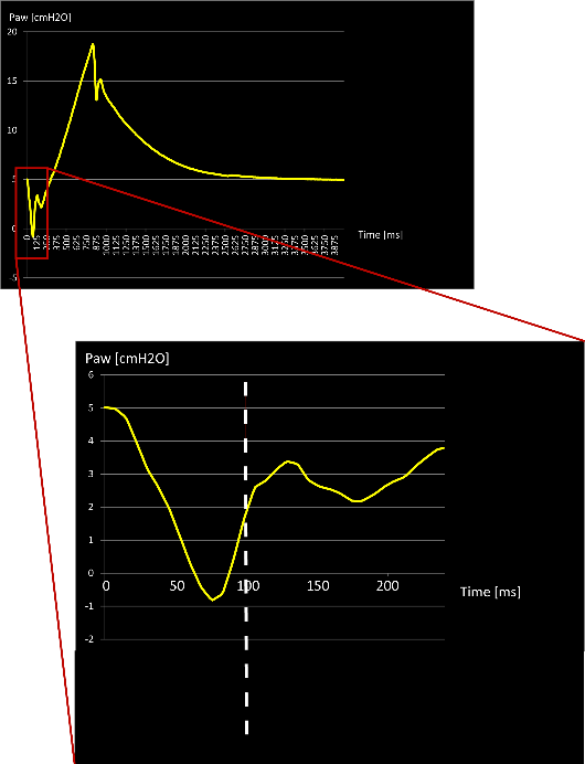
P0.1 corresponds to the drop in Paw, observed during the first 100 ms of an inspiratory effort made against the occluded airway opening (see Figure 1).

Current ICU ventilators maximize freedom for the patient. A fast response compensates pressure drops before 100 ms, especially when the inspiratory valves remain open to guarantee free breathing at any time (biphasic concept).
  • No occlusion
  • Pressure is compensated before the first 100 ms
Screenshot of ventilator display showing pressure drop
Figure 1
Screenshot of ventilator display showing pressure drop
Figure 1

Monitoring P0.1 with no occlusion maneuver

How can P0.1 be monitored without an occlusion maneuver?

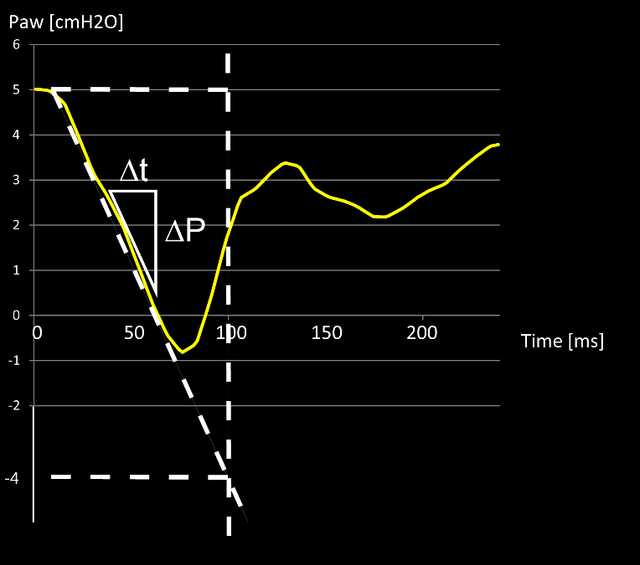
The ventilator calculates the steepest tangent of the drop in the pressure curve during an inspiratory effort.

This is shown in Figure 2 as a white triangle, and describes the minimum gradient of ∆t/∆P. 

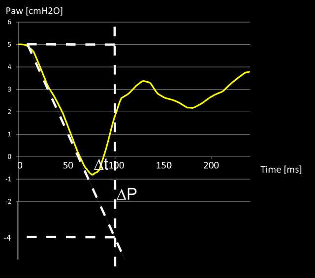
Once the minimum gradient is calculated, the value P0.1 is the extrapolation to 100 ms after pressure drops below PEEP.

Shown here in Figure 3 as a triangle with white dotted lines, P0.1 is the pressure drop below PEEP.

In this example:
P0.1 = Paw (100 ms) - PEEP = -9 cmH2O

P0.1 is monitored breath-by-breath, regardless of whether flow- or pressure-triggered (HAMILTON-G5/S1 pressure trigger only). 

Relevant devices: All

Full citations below: (Brenner M, Mukai DS, Russell JE, Spiritus EM, Wilson AF. A new method for measurement of airway occlusion pressure. Chest. 1990;98(2):421-427. doi:10.1378/chest.98.2.4211​)

Screenshot showing steepest tangent of drop in Paw as white triangle
Figure 2
Screenshot showing steepest tangent of drop in Paw as white triangle
Figure 2
Screenshot showing pressure drop below PEEP within first 100 ms marked with dotted line
Figure 3
Screenshot showing pressure drop below PEEP within first 100 ms marked with dotted line
Figure 3

A new method for measurement of airway occlusion pressure.

Brenner M, Mukai DS, Russell JE, Spiritus EM, Wilson AF. A new method for measurement of airway occlusion pressure. Chest. 1990;98(2):421-427. doi:10.1378/chest.98.2.421

Airway occlusion pressure correlates with central respiratory drive. The airway occlusion pressure (P0.1) may be an excellent predictor of the ability of patients with obstructive lung disease to wean from mechanical ventilation. We describe a new method for measuring P0.1 using digitized signals generated from standard respiratory equipment and a computer program to automatically determine P0.1 values. The accuracy of this new method was tested by comparison with standard analog recorder methods using a mechanical lung model, in ventilated patients in an intensive care unit, and in normal volunteers. In all settings, excellent correlation was obtained between P0.1 measurements by the digital Servo and standard analog methods (r = 0.99). This new method permits accurate and automatic determination of P0.1 in ventilated patients using standard respiratory equipment. The rapid response and ease of use of this method should enable evaluation of a number of physiologic variables involved in respiratory control in ventilated and nonventilated patients.